Mouse EGFR Antibody Summary
Leu25-Ser647
Accession # Q9EP98
Applications
Please Note: Optimal dilutions should be determined by each laboratory for each application. General Protocols are available in the Technical Information section on our website.
Scientific Data
 View Larger
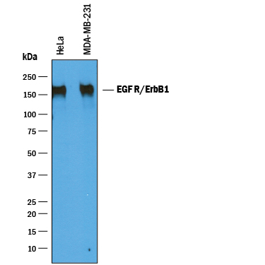
Detection of Human EGFR by Western Blot. Western blot shows lysates of HeLa human cervical epithelial carcinoma cell line and MDA-MB-231 human breast cancer cell line. PVDF membrane was probed with 0.25 µg/mL of Goat Anti-Mouse EGFR Antigen Affinity-purified Polyclonal Antibody (Catalog # AF1280) followed by HRP-conjugated Anti-Goat IgG Secondary Antibody (Catalog # HAF019). A specific band was detected for EGFR at approximately 170 kDa (as indicated). This experiment was conducted under reducing conditions and using Immunoblot Buffer Group 1.
 View Larger
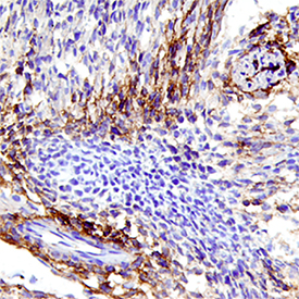
EGFR in Mouse Embryo. EGFR was detected in immersion fixed frozen sections of mouse embryo (13 d.p.c.) using Goat Anti-Mouse EGFR Antigen Affinity-purified Polyclonal Antibody (Catalog # AF1280) at 15 µg/mL overnight at 4 °C. Tissue was stained using the Anti-Goat HRP-DAB Cell & Tissue Staining Kit (brown; Catalog # CTS008) and counterstained with hematoxylin (blue). Specific staining was localized to developing muscle. View our protocol for Chromogenic IHC Staining of Frozen Tissue Sections.
 View Larger
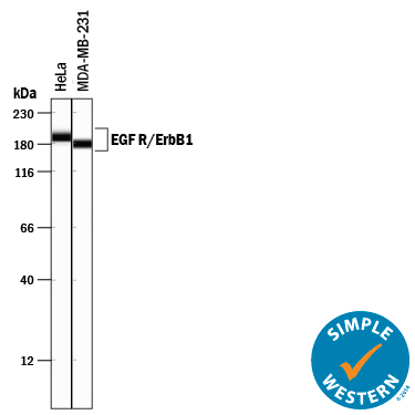
Detection of Human EGFR by Simple WesternTM. Simple Western lane view shows lysates of HeLa human cervical epithelial carcinoma cell line and MDA-MB-231 human breast cancer cell line, loaded at 0.2 mg/mL. A specific band was detected for EGFR at approximately 180-191 kDa (as indicated) using 2.5 µg/mL of Goat Anti-Mouse EGFR Antigen Affinity-purified Polyclonal Antibody (Catalog # AF1280) followed by 1:50 dilution of HRP-conjugated Anti-Goat IgG Secondary Antibody (Catalog # HAF109). This experiment was conducted under reducing conditions and using the 12-230 kDa separation system.
Reconstitution Calculator
Preparation and Storage
- 12 months from date of receipt, -20 to -70 °C as supplied.
- 1 month, 2 to 8 °C under sterile conditions after reconstitution.
- 6 months, -20 to -70 °C under sterile conditions after reconstitution.
Background: EGFR
The EGFR subfamily of receptor tyrosine kinases comprises four members: EGFR (also known as Her1, ErbB1, or ErbB), ErbB2 (Neu, Her2), ErbB3 (Her3), and ErbB4 (Her4). All family members are type I transmembrane glycoproteins. They contain an extracellular ligand binding domain containing two cysteine-rich domains and a cytoplasmic domain containing a membrane-proximal tyrosine kinase domain followed by multiple tyrosine autophosphorylation sites (1, 2). The mouse EGFR cDNA encodes a 1210 amino acid (aa) precursor with a 24 aa signal peptide, a 623 aa extracellular domain (ECD), a 23 aa transmembrane segment, and a 540 aa cytoplasmic domain (3). Soluble receptors consisting of the extracellular ligand binding domain are generated by alternate splicing in human and mouse (4-6). Within the ECD, mouse EGFR shares 88% and 93% aa sequence identity with human and rat EGFR, respectively. It shares 44-48% aa sequence identity with the ECD of mouse ErbB2, ErbB3, and ErbB4. EGFR binds a subset of the EGF family ligands, including EGF, amphiregulin, TGF-alpha, betacellulin, epiregulin, HB-EGF, and epigen (1, 2). Ligand binding induces EGFR homodimerization as well as heterodimerization with ErbB2, resulting in kinase activation, heterodimerization tyrosine phosphorylation and cell signaling (7-11). EGFR can also be recruited to form heterodimers with the ligand-activated ErbB3 or ErbB4. EGFR signaling regulates multiple biological functions including cell proliferation, differentiation, motility, and apoptosis (12, 13). EGFR is over-expressed in a wide variety of tumors and is the target of several anti-cancer drugs (14).
- Singh, A.B. and R.C. Harris (2005) Cell. Signal. 17:1183.
- Shilo, B.Z. (2005) Development 132:4017.
- Avivi, A. et al. (1991) Oncogene 6:673.
- Reiter, J.L. and N.J. Maihle (1996) Nucleic Acids Res. 24:4050.
- Reiter J.L. et al. (2001) Genomics 71:1.
- Xu, Y.H. et al. (1984) Nature 309:806.
- Graus-Porta, D. et al. (1997) EMBO J. 16:1647.
- Yarden, Y. et al. (1987) Biochemistry 26:1434.
- Burgess, A.W. et al. (2003) Mol. Cell 12:541.
- Lemmon, M.A. et al. (1997) EMBO J. 16:281.
- Cohen, S. et al. (1982) J. Biol. Chem. 257:1523.
- Sibilia, M. and E.F. Wagner (1995) Science 269:234.
- Miettinen, P.J. et al. (1995) Nature 376:337.
- Roskoski Jr., R. (2004) Biochem. Biophys. Res. Commun. 319:1.
Product Datasheets
Citations for Mouse EGFR Antibody
R&D Systems personnel manually curate a database that contains references using R&D Systems products. The data collected includes not only links to publications in PubMed, but also provides information about sample types, species, and experimental conditions.
8
Citations: Showing 1 - 8
Filter your results:
Filter by:
-
Variation of Human Neural Stem Cells Generating Organizer States In�Vitro before Committing to Cortical Excitatory or Inhibitory Neuronal Fates
Authors: N Micali, SK Kim, M Diaz-Busta, G Stein-O'Br, S Seo, JH Shin, BG Rash, S Ma, Y Wang, NA Olivares, JI Arellano, KR Maynard, EJ Fertig, AJ Cross, RW Bürli, NJ Brandon, DR Weinberger, JG Chenoweth, DJ Hoeppner, N Sestan, P Rakic, C Colantuoni, RD McKay
Cell Rep, 2020-05-05;31(5):107599.
Species: Human
Sample Types:
Applications: IF -
Nanobody-targeting of epidermal growth factor receptor (EGFR) ectodomain variants overcomes resistance to therapeutic EGFR antibodies
Authors: J Tintelnot, N Baum, C Schultheis, F Braig, M Trentmann, J Finter, W Fumey, P Bannas, B Fehse, K Riecken, K Schütze, C Bokemeyer, T Rösner, T Valerius, M Peipp, F Koch-Nolte, M Binder
Mol. Cancer Ther., 2019-03-01;0(0):.
Species: Mouse
Sample Types: Whole Cells
Applications: Flow Cytometry -
Epidermal growth factor receptor promotes cerebral and retinal invasion by Toxoplasma gondii
Authors: YL Corcino, JC Portillo, CS Subauste
Sci Rep, 2019-01-24;9(1):669.
Species: Mouse
Sample Types: Cell Lysates
Applications: Western Blot -
The Immunogenicity and Anti-Tumor Efficacy of a Rationally Designed EGFR Vaccine
Authors: C Cheng, L Deng, R Li
Cell. Physiol. Biochem., 2018-03-20;46(1):46-56.
Species: Mouse
Sample Types: Protein
Applications: Western Blot -
iRhom2 promotes lupus nephritis through TNF-? and EGFR signaling
Authors: X Qing, Y Chinenov, P Redecha, M Madaio, JJ Roelofs, G Farber, PD Issuree, L Donlin, DR McIlwain, TW Mak, CP Blobel, JE Salmon
J. Clin. Invest., 2018-03-05;0(0):.
Species: Mouse
Sample Types: Tissue Homogenates
Applications: Western Blot -
Endocannabinoid Signaling in Embryonic Neuronal Motility and Cell-Cell Contact - Role of mGluR5 and TRPC3 Channels
Authors: PM Turunen, LM Louhivuori, V Louhivuori, JP Kukkonen, KE Åkerman
Neuroscience, 2018-02-10;0(0):.
Species: Mouse
Sample Types: Whole Cells
Applications: ICC -
EGF transactivation of Trk receptors regulates the migration of newborn cortical neurons.
Authors: Puehringer D, Orel N, Luningschror P, Subramanian N, Herrmann T, Chao M, Sendtner M
Nat Neurosci, 2013-02-17;16(4):407-15.
Species: Mouse
Sample Types: Tissue Homogenates
Applications: Immunoprecipitation -
T cells enhance stem-like properties and conditional malignancy in gliomas.
Authors: Irvin DK, Jouanneau E, Duvall G
PLoS ONE, 2010-06-07;5(6):e10974.
Species: Mouse
Sample Types: Whole Cells
Applications: Flow Cytometry
FAQs
No product specific FAQs exist for this product, however you may
View all Antibody FAQsReviews for Mouse EGFR Antibody
There are currently no reviews for this product. Be the first to review Mouse EGFR Antibody and earn rewards!
Have you used Mouse EGFR Antibody?
Submit a review and receive an Amazon gift card.
$25/€18/£15/$25CAN/¥75 Yuan/¥2500 Yen for a review with an image
$10/€7/£6/$10 CAD/¥70 Yuan/¥1110 Yen for a review without an image


