Mouse FOLR1 Antibody Summary
Arg23-Ser232
Accession # P35846
Applications
Please Note: Optimal dilutions should be determined by each laboratory for each application. General Protocols are available in the Technical Information section on our website.
Scientific Data
 View Larger
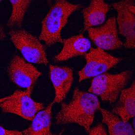
FOLR1 in HeLa Human Cell Line. FOLR1 was detected in immersion fixed HeLa human cervical epithelial carcinoma cell line using Sheep Anti-Mouse FOLR1 Antigen Affinity-purified Polyclonal Antibody (Catalog # AF6936) at 10 µg/mL for 3 hours at room temperature. Cells were stained using the NorthernLights™ 557-conjugated Anti-Sheep IgG Secondary Antibody (red; Catalog # NL010) and counterstained with DAPI (blue). Specific staining was localized to cytoplasm. View our protocol for Fluorescent ICC Staining of Cells on Coverslips.
 View Larger
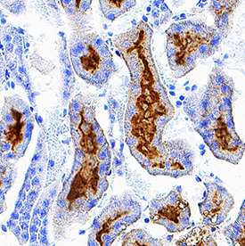
FOLR1 in Mouse Kidney. FOLR1 was detected in perfusion fixed frozen sections of mouse kidney using Sheep Anti-Mouse FOLR1 Antigen Affinity-purified Polyclonal Antibody (Catalog # AF6936) at 0.3 µg/mL overnight at 4 °C. Tissue was stained using the Anti-Sheep HRP-DAB Cell & Tissue Staining Kit (brown; Catalog # CTS019) and counterstained with hematoxylin (blue). Specific staining was localized to convoluted tubules. View our protocol for Chromogenic IHC Staining of Frozen Tissue Sections.
Reconstitution Calculator
Preparation and Storage
- 12 months from date of receipt, -20 to -70 °C as supplied.
- 1 month, 2 to 8 °C under sterile conditions after reconstitution.
- 6 months, -20 to -70 °C under sterile conditions after reconstitution.
Background: FOLR1
Folate receptor 1 (FOLR1; also folate receptor alpha and folate binding protein 1/FOLBP1) is a 40-50 kDa member of the folate receptor family. FOLR1 is expressed in reticulocytes, retinal Muller cells and placenta, plus renal, mammary and thymic epithelium, where it mediates the transport of 5-methyltetrahydrofolate into the cell. The mouse FOLR1 preproprecursor is 255 amino acid (aa) in length. It contains a GPI-linked glycoprotein that is 208 aa in size (aa 25-232), and which possesses three potential N-linked glycosylation sites. There is one potential isoform variant that shows a 105 aa substitution for aa 110-255. Over aa 23-232, mouse FOLR1 shares 83% and 93% aa sequence identity with human and rat FOLR1, respectively.
Product Datasheets
Citation for Mouse FOLR1 Antibody
R&D Systems personnel manually curate a database that contains references using R&D Systems products. The data collected includes not only links to publications in PubMed, but also provides information about sample types, species, and experimental conditions.
1 Citation: Showing 1 - 1
-
Reinforcing one-carbon metabolism via folic acid/Folr1 promotes &beta-cell differentiation
Authors: C Karampelia, H Rezanejad, M Rosko, L Duan, J Lu, L Pazzagli, P Bertolino, CE Cesta, X Liu, GS Korbutt, O Andersson
Nature Communications, 2021-06-07;12(1):3362.
Species: Mouse
Sample Types: Whole Tissue
Applications: IHC
FAQs
No product specific FAQs exist for this product, however you may
View all Antibody FAQsReviews for Mouse FOLR1 Antibody
Average Rating: 5 (Based on 1 Review)
Have you used Mouse FOLR1 Antibody?
Submit a review and receive an Amazon gift card.
$25/€18/£15/$25CAN/¥75 Yuan/¥2500 Yen for a review with an image
$10/€7/£6/$10 CAD/¥70 Yuan/¥1110 Yen for a review without an image
Filter by:


