Mouse IL-1 beta /IL-1F2 Antibody Summary
Val118-Ser269
Accession # P10749
Applications
Please Note: Optimal dilutions should be determined by each laboratory for each application. General Protocols are available in the Technical Information section on our website.
Scientific Data
 View Larger
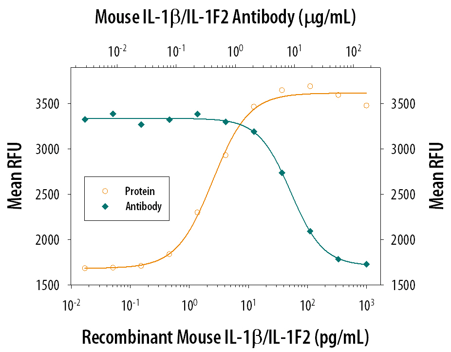
Cell Proliferation Induced by IL‑1 beta /IL‑1F2 and Neutral-ization by Mouse IL‑1 beta /IL‑1F2 Antibody. Recombinant Mouse IL-1 beta /IL-1F2 (401-ML) stimulates proliferation in the the D10.G4.1 mouse helper T cell line in a dose-dependent manner (orange line). Proliferation elicited by Recombinant Mouse IL-1 beta /IL-1F2 (10 pg/mL) is neutralized (green line) by increasing concentrations of Goat Anti-Mouse IL-1 beta /IL-1F2 Polyclonal Antibody (Catalog # AB-401-NA). The ND50 is typically 2-12 µg/mL.
 View Larger
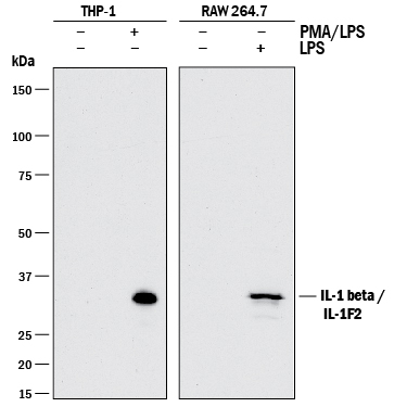
Detection of Human and Mouse IL‑1 beta /IL‑1F2 by Western Blot. Western blot shows lysates of THP-1 human acute monocytic leukemia cell line untreated (-) or treated (+) with 200 nM PMA for 24 hours and 10 µg/mL LPS for 4 hours and RAW 264.7 mouse monocyte/macrophage cell line untreated (-) or treated (+) with 10 µg/mL LPS for 24 hours. PVDF membrane was probed with 1 µg/mL of Goat Anti-Mouse IL-1 beta /IL-1F2 Polyclonal Antibody (Catalog # AB-401-NA) followed by HRP-conjugated Anti-Goat IgG Secondary Antibody (HAF017). A specific band was detected for IL-1 beta /IL-1F2 at approximately 35 kDa (as indicated). This experiment was conducted under reducing conditions and using Immunoblot Buffer Group 1.
 View Larger
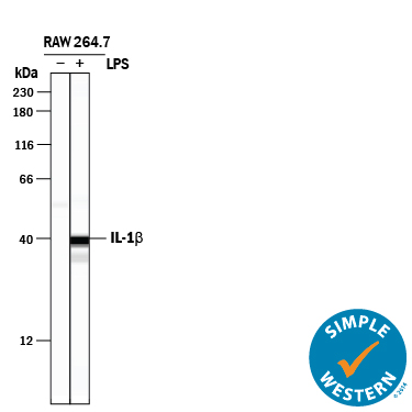
Detection of Mouse IL‑1 beta /IL‑1F2 by Simple WesternTM. Simple Western lane view shows lysates of RAW 264.7 mouse monocyte/macrophage cell line untreated (-) or treated (+) with 10 µg/mL LPS for 24 hours, loaded at 0.5 mg/mL. A specific band was detected for IL‑1 beta /IL‑1F2 at approximately 40 kDa (as indicated) using 50 µg/mL of Goat Anti-Mouse IL‑1 beta /IL‑1F2 Polyclonal Antibody (Catalog # AB-401-NA). This experiment was conducted under reducing conditions and using the 12-230 kDa separation system.
Reconstitution Calculator
Preparation and Storage
- 12 months from date of receipt, -20 to -70 °C as supplied.
- 1 month, 2 to 8 °C under sterile conditions after reconstitution.
- 6 months, -20 to -70 °C under sterile conditions after reconstitution.
Background: IL-1 beta/IL-1F2
IL-1 is a name that designates two pleiotropic cytokines, IL-1 alpha (IL-1F1) and IL-1 beta (IL-1F2, IL1B), which are the products of distinct genes. IL-1 alpha and IL-1 beta are structurally related polypeptides that share approximately 17% amino acid (aa) identity in mouse. Both proteins are produced by a wide variety of cells in response to inflammatory agents, infections, or microbial endotoxins. While IL-1 alpha and IL-1 beta are regulated independently, they bind to the same receptor and exert identical biological effects. IL-1 RI binds directly to IL-1 alpha or IL-1 beta and then associates with IL-1 R accessory protein (IL-1 R3/IL-1 R AcP) to form a high-affinity receptor complex that is competent for signal transduction. IL-1 RII has high affinity for IL-1 beta but functions as a decoy receptor and negative regulator of IL-1 beta activity. IL-1ra functions as a competitive antagonist by preventing IL-1 alpha and IL-1 beta from interacting with IL-1 RI. Intracellular cleavage of the IL-1 beta precursor by Caspase-1/ICE is a key step in the inflammatory response. The 17 kDa molecular weight mature mouse IL-1 beta shares 90% aa sequence identity with cotton rat and rat and 67%-78% with canine, equine, feline, human, porcine, and rhesus macaque IL-1 beta. IL-1 beta functions in a central role in immune and inflammatory responses, bone remodeling, fever, carbohydrate metabolism, and GH/IGF-I physiology. IL-1 beta dysregulation is implicated in many pathological conditions including sepsis, rheumatoid arthritis, inflammatory bowel disease, acute and chronic myelogenous leukemia, insulin-dependent diabetes mellitus, atherosclerosis, neuronal injury, and aging-related diseases.
Product Datasheets
Citations for Mouse IL-1 beta /IL-1F2 Antibody
R&D Systems personnel manually curate a database that contains references using R&D Systems products. The data collected includes not only links to publications in PubMed, but also provides information about sample types, species, and experimental conditions.
16
Citations: Showing 1 - 10
Filter your results:
Filter by:
-
The inflammatory microenvironment of the lung at the time of infection governs innate control of SARS-CoV-2 replication
Authors: Baker, PJ;Bohrer, AC;Castro, E;Amaral, EP;Snow-Smith, M;Torres-Juárez, F;Gould, ST;Queiroz, ATL;Fukutani, ER;Jordan, CM;Khillan, JS;Cho, K;Barber, DL;Andrade, BB;Johnson, RF;Hilligan, KL;Mayer-Barber, KD;
bioRxiv : the preprint server for biology
Species: Mouse
Sample Types: Cell Lysates
Applications: Western Blot -
N6-Methyladenosine and Reader Protein YTHDF2 Enhance the Innate Immune Response by Mediating DUSP1 mRNA Degradation and Activating Mitogen-Activated Protein Kinases during Bacterial and Viral Infections
Authors: J Feng, W Meng, L Chen, X Zhang, A Markazi, W Yuan, Y Huang, SJ Gao
MBio, 2023-01-10;0(0):e0334922.
Species: Mouse
Sample Types: Cell Lysates
Applications: Western Blot -
Repositioning of the Angiotensin II Receptor Antagonist Candesartan as an Anti-Inflammatory Agent With NLRP3 Inflammasome Inhibitory Activity
Authors: Lin WY, Li LH, Hsiao YY et al.
Frontiers in Immunology
-
GSK3beta mediates the spatiotemporal dynamics of NLRP3 inflammasome activation
Authors: S Arumugam, Y Qin, Z Liang, SN Han, SLT Boodapati, J Li, Q Lu, RA Flavell, WZ Mehal, X Ouyang
Cell Death and Differentiation, 2022-04-27;0(0):.
Species: Mouse
Sample Types: Cell Culture Supernates
Applications: Western Blot -
Assessing the Association of Mitochondrial Function and Inflammasome Activation in Murine Macrophages Exposed to Select Mitotoxic Tri-Organotin Compounds
Authors: GM Childers, CA Perry, B Blachut, N Martin, CD Bortner, S Sieber, JL Li, MB Fessler, GJ Harry
Environmental health perspectives, 2021-04-30;129(4):47015.
Species: Mouse
Sample Types: Cell Lysates
Applications: Western Blot -
A Synthetic Small Molecule F240B Decreases NLRP3 Inflammasome Activation by Autophagy Induction
Authors: Wu CH, Gan CH, Li LH et al.
Frontiers in Immunology
-
The protective role of proton-sensing TDAG8 in the brain injury in a mouse ischemia reperfusion model
Authors: K Sato, A Tobo, C Mogi, M Tobo, N Yamane, M Tosaka, H Tomura, DS Im, F Okajima
Sci Rep, 2020-10-14;10(1):17193.
Species: Mouse
Sample Types: Cell Lysates
Applications: Western Blot -
Mice Lacking the Purinergic Receptor P2X5 Exhibit Defective Inflammasome Activation and Early Susceptibility to Listeria monocytogenes
Authors: YH Jeong, MC Walsh, J Yu, H Shen, EJ Wherry, Y Choi
J. Immunol., 2020-06-15;0(0):.
Species: Mouse
Sample Types: Cell Lysates
Applications: Western Blot -
Coenzyme Q10 protects against burn-induced mitochondrial dysfunction and impaired insulin signaling in mouse skeletal muscle
Authors: H Nakazawa, K Ikeda, S Shinozaki, S Yasuhara, YM Yu, JAJ Martyn, RG Tompkins, T Yorozu, S Inoue, M Kaneki
FEBS Open Bio, 2019-01-19;9(2):348-363.
Species: Mouse
Sample Types: Tissue Homogenates
Applications: Western Blot -
PtdIns4P on dispersed trans-Golgi network mediates NLRP3 inflammasome activation
Authors: J Chen, ZJ Chen
Nature, 2018-11-28;564(7734):71-76.
Species: Mouse
Sample Types: Cell Lysates
Applications: Western Blot -
P2RX7 sensitizes Mac-1/ICAM-1-dependent leukocyte-endothelial adhesion and promotes neurovascular injury during septic encephalopathy.
Authors: Wang H, Hong L, Huang J, Jiang Q, Tao R, Tan C, Lu N, Wang C, Ahmed M, Lu Y, Liu Z, Shi W, Lai E, Wilcox C, Han F
Cell Res, 2015-05-22;25(6):674-90.
Species: Mouse
Sample Types: In Vivo
Applications: Neutralization -
A small-molecule inhibitor of the NLRP3 inflammasome for the treatment of inflammatory diseases.
Authors: Coll R, Robertson A, Chae J, Higgins S, Munoz-Planillo R, Inserra M, Vetter I, Dungan L, Monks B, Stutz A, Croker D, Butler M, Haneklaus M, Sutton C, Nunez G, Latz E, Kastner D, Mills K, Masters S, Schroder K, Cooper M, O'Neill L
Nat Med, 2015-02-16;21(3):248-55.
Species: Mouse
Sample Types: Cell Lysates
Applications: Western Blot -
Distinct mechanisms of induction of hepatic growth hormone resistance by endogenous IL-6, TNF-alpha, and IL-1beta.
Authors: Zhao Y, Xiao X, Frank S, Lin H, Xia Y
Am J Physiol Endocrinol Metab, 2014-06-03;307(2):E186-98.
Species: Mouse
Sample Types: In Vivo
Applications: Neutralization -
Interleukin-1beta and fibroblast growth factor receptor 1 cooperate to induce cyclooxygenase-2 during early mammary tumourigenesis.
Authors: Reed JR, Leon RP, Hall MK, Schwertfeger KL
Breast Cancer Res., 2009-04-24;11(2):R21.
Species: Mouse
Sample Types: In Vivo
Applications: Neutralization -
Comparison of the roles of IL-1, IL-6, and TNFalpha in cell culture and murine models of aseptic loosening.
Authors: Taki N, Tatro JM, Lowe R, Goldberg VM, Greenfield EM
Bone, 2006-12-21;40(5):1276-83.
Species: Mouse
Sample Types: Whole Cells
Applications: Neutralization -
The Myc-dependent angiogenic switch in tumors is mediated by interleukin 1beta.
Authors: Shchors K, Shchors E, Rostker F, Lawlor ER, Brown-Swigart L, Evan GI
Genes Dev., 2006-09-15;20(18):2527-38.
Species: Mouse
Sample Types: In Vivo
Applications: Neutralization
FAQs
No product specific FAQs exist for this product, however you may
View all Antibody FAQsReviews for Mouse IL-1 beta /IL-1F2 Antibody
There are currently no reviews for this product. Be the first to review Mouse IL-1 beta /IL-1F2 Antibody and earn rewards!
Have you used Mouse IL-1 beta /IL-1F2 Antibody?
Submit a review and receive an Amazon gift card.
$25/€18/£15/$25CAN/¥75 Yuan/¥2500 Yen for a review with an image
$10/€7/£6/$10 CAD/¥70 Yuan/¥1110 Yen for a review without an image


