Mouse LAG-3 PE-conjugated Antibody Summary
Ser23-Leu442
Accession # Q61790
Applications
Please Note: Optimal dilutions should be determined by each laboratory for each application. General Protocols are available in the Technical Information section on our website.
Scientific Data
 View Larger
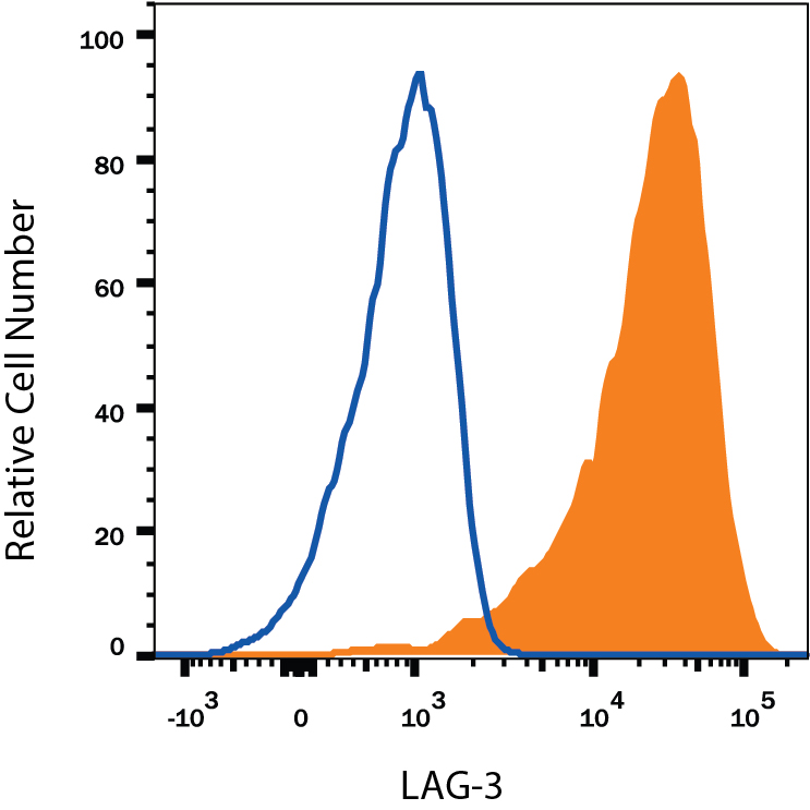
Detection of LAG-3 in Mouse Splenocytes by Flow Cytometry. Mouse splenocytes treated with PMA and Calcium Ionomycin for 3 days were stained with Rat Anti-Mouse LAG-3 PE-conjugated Monoclonal Antibody (Catalog # FAB33281P, filled histogram) or isotype control antibody (Catalog # IC013P, open histogram). View our protocol for Staining Membrane-associated Proteins.
Reconstitution Calculator
Preparation and Storage
- 12 months from date of receipt, 2 to 8 °C as supplied.
Background: LAG-3
LAG-3 (Lymphocyte activation gene-3; also known as CD223 in the human) is a member of the immunoglobulin superfamily (IgSF). The mature LAG-3 protein is a 496 amino acid (aa) membrane protein with a 421 aa extracellular region which contains four IgSF domains, a 21 aa transmembrane region and a 54 aa cytoplasmic region. LAG-3 shares < 20% amino acid sequence homology with CD4, but has similar structure and binds to MHC class II with higher affinity. The mouse LAG-3 extracellular region shares 69% aa sequence identity with human LAG-3.
Product Datasheets
FAQs
No product specific FAQs exist for this product, however you may
View all Antibody FAQsReviews for Mouse LAG-3 PE-conjugated Antibody
There are currently no reviews for this product. Be the first to review Mouse LAG-3 PE-conjugated Antibody and earn rewards!
Have you used Mouse LAG-3 PE-conjugated Antibody?
Submit a review and receive an Amazon gift card.
$25/€18/£15/$25CAN/¥75 Yuan/¥2500 Yen for a review with an image
$10/€7/£6/$10 CAD/¥70 Yuan/¥1110 Yen for a review without an image

