Mouse/Rat Cathepsin C/DPPI Antibody Summary
Asp25-Leu462
Accession # P97821
Applications
Please Note: Optimal dilutions should be determined by each laboratory for each application. General Protocols are available in the Technical Information section on our website.
Scientific Data
 View Larger
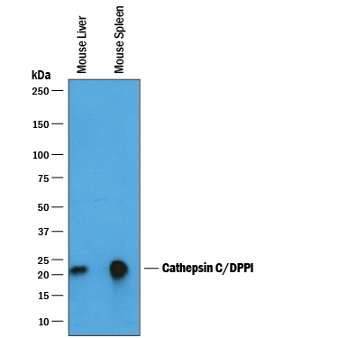
Detection of Mouse Cathepsin C/DPPI by Western Blot. Western blot shows lysates of mouse liver tissue and mouse spleen tissue. PVDF membrane was probed with 0.25 µg/mL of Goat Anti-Mouse Cathepsin C/DPPI Antigen Affinity-purified Polyclonal Antibody (Catalog # AF1034) followed by HRP-conjugated Anti-Goat IgG Secondary Antibody (Catalog # HAF019). A specific band was detected for Cathepsin C/DPPI at approximately 20-25 kDa (as indicated). This experiment was conducted under reducing conditions and using Immunoblot Buffer Group 1.
 View Larger
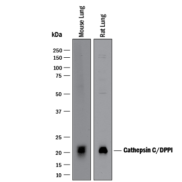
Detection of Mouse and Rat Cathepsin C/DPPI by Western Blot. Western blot shows lysates of mouse lung tissue and rat lung tissue. PVDF membrane was probed with 1 µg/mL of Goat Anti-Mouse Cathepsin C/DPPI Antigen Affinity-purified Polyclonal Antibody (Catalog # AF1034) followed by HRP-conjugated Anti-Goat IgG Secondary Antibody (Catalog # HAF017). A specific band was detected for Cathepsin C/DPPI at approximately 20-22 kDa (as indicated). This experiment was conducted under reducing conditions and using Immunoblot Buffer Group 1.
 View Larger
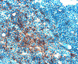
Cathepsin C/DPPI in Mouse Thymus. Cathepsin C/DPPI was detected in perfusion fixed frozen sections of mouse thymus using Goat Anti-Mouse Cathepsin C/ DPPI Antigen Affinity-purified Polyclonal Antibody (Catalog # AF1034) at 15 µg/mL overnight at 4 °C. Tissue was stained using the Anti-Goat HRP-DAB Cell & Tissue Staining Kit (brown; Catalog # CTS008) and counter-stained with hematoxylin (blue). Specific staining was localized to thymocytes. View our protocol for Chromogenic IHC Staining of Frozen Tissue Sections.
 View Larger
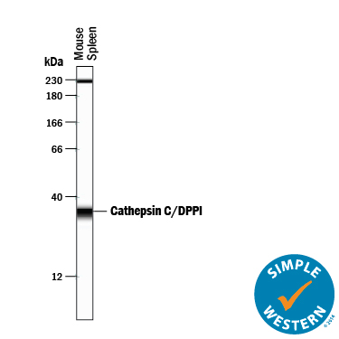
Detection of Mouse Cathepsin C/DPPI by Simple WesternTM. Simple Western lane view shows lysates of mouse spleen tissue, loaded at 0.2 mg/mL. A specific band was detected for Cathepsin C/DPPI at approximately 35 kDa (as indicated) using 2.5 µg/mL of Goat Anti-Mouse Cathepsin C/DPPI Antigen Affinity-purified Polyclonal Antibody (Catalog # AF1034) followed by 1:50 dilution of HRP-conjugated Anti-Goat IgG Secondary Antibody (Catalog # HAF109). This experiment was conducted under reducing conditions and using the 12-230 kDa separation system. Non-specific interaction with the 230 kDa Simple Western standard may be seen with this antibody.
Reconstitution Calculator
Preparation and Storage
- 12 months from date of receipt, -20 to -70 °C as supplied.
- 1 month, 2 to 8 °C under sterile conditions after reconstitution.
- 6 months, -20 to -70 °C under sterile conditions after reconstitution.
Background: Cathepsin C/DPPI
Cathepsin C is a cysteine protease of the papain family (1). Cathepsin C sequentially removes dipeptides from the free N-termini of proteins and peptides. It has broad specificity except that it does not cleave a basic amino acid (Arg or Lys) in the N-terminal position or Pro on either side of the scissle bond. It requires halide ions for activity. The pro form contains a pro peptide and a catalytic region, which can be further processed into heavy/ alpha and light/ beta chains that are linked by a disulfide bond. It is broadly distributed. Cathepsin C plays a role in the lysosomal degradation. It also functions as a key enzyme in the activation of granule serine proteases in cytotoxic T lymphocytes and natural killer cells (granzymes A and B), mast cells (tryptase and chymase), and neutrophils (Cathepsin G and elastase) by removing their N-terminal activation dipeptides (2).
- Turk, B. et al. (2004) in Handbook of Proteolytic Enzymes. Barrett, et al. eds. p. 1192, Academic Press, San Diego.
- Dahl, S.W. et al. (2001) Biochemistry 40:1671.
Product Datasheets
Citations for Mouse/Rat Cathepsin C/DPPI Antibody
R&D Systems personnel manually curate a database that contains references using R&D Systems products. The data collected includes not only links to publications in PubMed, but also provides information about sample types, species, and experimental conditions.
10
Citations: Showing 1 - 10
Filter your results:
Filter by:
-
Inflammatory Serine Proteases Play a Critical Role in the Early Pathogenesis of Diabetic Cardiomyopathy
Authors: MA Kolpakov, K Sikder, A Sarkar, S Chaki, SK Shukla, X Guo, Z Qi, C Barbery, A Sabri, K Rafiq
Cell. Physiol. Biochem., 2019-01-01;53(6):982-998.
Species: Mouse
Sample Types: Whole Tissue
Applications: IHC-P -
The balance between cathepsin C and cystatin F controls remyelination in the brain of Plp1-overexpressing mouse, a chronic demyelinating disease model
Authors: T Shimizu, W Wisessmith, J Li, M Abe, K Sakimura, B Chetsawang, Y Sahara, K Tohyama, KF Tanaka, K Ikenaka
Glia, 2017-03-02;0(0):.
Species: Mouse
Sample Types: Whole Cells, Whole Tissue
Applications: ICC, IHC -
Deficiency for the cysteine protease cathepsin L impairs Myc-induced tumorigenesis in a mouse model of pancreatic neuroendocrine cancer.
Authors: Brindle N, Joyce J, Rostker F, Lawlor E, Swigart-Brown L, Evan G, Hanahan D, Shchors K
PLoS ONE, 2015-04-30;10(4):e0120348.
Species: Mouse
Sample Types: Whole Tissue
Applications: IHC -
Lysosomal protein turnover contributes to the acquisition of TGFbeta-1 induced invasive properties of mammary cancer cells.
Authors: Kern U, Wischnewski V, Biniossek M, Schilling O, Reinheckel T
Mol Cancer, 2015-02-15;14(0):39.
Species: Mouse
Sample Types: Cell Lysates
Applications: Western Blot -
The endolysosomal cysteine cathepsins L and K are involved in macrophage-mediated clearance of Staphylococcus aureus and the concomitant cytokine induction.
Authors: Muller S, Faulhaber A, Sieber C, Pfeifer D, Hochberg T, Gansz M, Deshmukh S, Dauth S, Brix K, Saftig P, Peters C, Henneke P, Reinheckel T
FASEB J, 2013-09-13;28(1):162-75.
Species: Mouse
Sample Types: Cell Lysates
Applications: Western Blot -
Up-regulation of microglial cathepsin C expression and activity in lipopolysaccharide-induced neuroinflammation.
Authors: Fan K, Wu X, Fan B, Li N, Lin Y, Yao Y, Ma J
J Neuroinflammation, 2012-05-20;9(1):96.
Species: Human
Sample Types: Whole Tissue
Applications: IHC -
Macrophages and cathepsin proteases blunt chemotherapeutic response in breast cancer.
Authors: Shree T, Olson OC, Elie BT
Genes Dev., 2011-12-01;25(23):2465-79.
Species: Mouse
Sample Types: Tissue Homogenates
Applications: Western Blot -
Gene targeting of the cysteine peptidase cathepsin H impairs lung surfactant in mice.
Authors: Buhling F, Kouadio M, Chwieralski CE, Kern U, Hohlfeld JM, Klemm N, Friedrichs N, Roth W, Deussing JM, Peters C, Reinheckel T
PLoS ONE, 2011-10-12;6(10):e26247.
Species: Mouse
Sample Types: Tissue Homogenates
Applications: Western Blot -
Mice deficient in LMAN1 exhibit FV and FVIII deficiencies and liver accumulation of {alpha}1-antitrypsin.
Authors: Zhang B, Zheng C, Zhu M, Tao J, Vasievich MP, Baines A, Kim J, Schekman R, Kaufman RJ, Ginsburg D
Blood, 2011-07-27;118(12):3384-91.
Species: Mouse
Sample Types: Whole Cells
Applications: ICC -
A role for serglycin proteoglycan in mast cell apoptosis induced by a secretory granule-mediated pathway.
Authors: Melo FR, Waern I, Ronnberg E, Abrink M, Lee DM, Schlenner SM, Feyerabend TB, Rodewald HR, Turk B, Wernersson S, Pejler G
J. Biol. Chem., 2010-12-01;286(7):5423-33.
Species: Mouse
Sample Types: Cell Lysates
Applications: Western Blot
FAQs
No product specific FAQs exist for this product, however you may
View all Antibody FAQsReviews for Mouse/Rat Cathepsin C/DPPI Antibody
Average Rating: 3 (Based on 1 Review)
Have you used Mouse/Rat Cathepsin C/DPPI Antibody?
Submit a review and receive an Amazon gift card.
$25/€18/£15/$25CAN/¥75 Yuan/¥2500 Yen for a review with an image
$10/€7/£6/$10 CAD/¥70 Yuan/¥1110 Yen for a review without an image
Filter by:

