Mouse/Rat Notch-1 Antibody Summary
Arg20-Glu488 (Ala208Thr, Asp334Glu)
Accession # Q07008
Applications
Please Note: Optimal dilutions should be determined by each laboratory for each application. General Protocols are available in the Technical Information section on our website.
Scientific Data
 View Larger
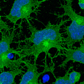
Notch‑1 in Rat Cortical Stem Cells. Notch-1 was detected in immersion fixed undifferentiated rat cortical stem cells using Goat Anti-Mouse/Rat Notch-1 Antigen Affinity-purified Polyclonal Antibody (Catalog # AF1057) at 10 µg/mL for 3 hours at room temperature. Cells were stained using the NorthernLights™ 493-conjugated Anti-Goat IgG Secondary Antibody (green; Catalog # NL003) and counterstained with DAPI (blue). Specific staining was localized to cell surfaces. View our protocol for Fluorescent ICC Staining of Stem Cells on Coverslips.
 View Larger
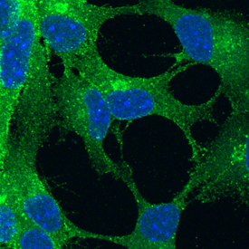
Notch‑1 in Mouse Cortical Stem Cells. Notch-1 was detected in immersion fixed undifferentiated mouse cortical stem cells using Goat Anti-Mouse/Rat Notch-1 Antigen Affinity-purified Polyclonal Antibody (Catalog # AF1057) at 10 µg/mL for 3 hours at room temperature. Cells were stained using the NorthernLights™ 493-conjugated Anti-Goat IgG Secondary Antibody (green; Catalog # NL003) and counterstained with DAPI (blue). Specific staining was localized to cell surfaces. View our protocol for Fluorescent ICC Staining of Stem Cells on Coverslips.
 View Larger
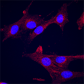
Notch‑1 in C2C12 Mouse Cell Line. Notch‑1 was detected in immersion fixed C2C12 mouse myoblast cell line using Goat Anti-Mouse/Rat Notch‑1 Antigen Affinity-purified Polyclonal Antibody (Catalog # AF1057) at 15 µg/mL for 3 hours at room temperature. Cells were stained using the NorthernLights™ 557-conjugated Anti-Goat IgG Secondary Antibody (red; NL001) and counterstained with DAPI (blue). Specific staining was localized to cytoplasm. Staining was performed using our protocol for Fluorescent ICC Staining of Non-adherent Cells.
 View Larger
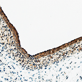
Notch‑1 in Rat Embryo. Notch-1 was detected in immersion fixed paraffin-embedded sections of rat embryo (13 d.p.c.) using Goat Anti-Mouse/Rat Notch-1 Antigen Affinity-purified Polyclonal Antibody (Catalog # AF1057) at 10 µg/mL overnight at 4 °C. Tissue was stained using the Anti-Goat HRP-DAB Cell & Tissue Staining Kit (brown; Catalog # CTS008) and counterstained with hematoxylin (blue). Specific staining was localized to labeling is in epidermis. View our protocol for Chromogenic IHC Staining of Paraffin-embedded Tissue Sections.
Reconstitution Calculator
Preparation and Storage
- 12 months from date of receipt, -20 to -70 °C as supplied.
- 1 month, 2 to 8 °C under sterile conditions after reconstitution.
- 6 months, -20 to -70 °C under sterile conditions after reconstitution.
Background: Notch-1
Rat Notch-1 is a 300 kDa, type I transmembrane glycoprotein involved in a number of early-event developmental processes (1). In both vertebrates and invertebrates, Notch signaling is important for specifying cell fates and for defining boundaries between different cell types. The molecule is synthesized as a 2531 amino acid (aa) precursor that contains an 18 aa signal sequence, a 1705 aa extracellular region, a 23 aa transmembrane (TM) segment and a 785 aa cytoplasmic domain (2). The large Notch-1 extracellular domain has 36 EGF‑like repeats followed by three notch/Lin-12 repeats. Of the 36 EGF‑like repeats, the 11th and 12th EGF‑like repeats have been shown to be both necessary and sufficient for binding the ligands Delta and Serrate, in Drosophila (3). The Notch-1 cytoplasmic domain contains six ankyrin repeats, a glutamine-rich domain and a PEST sequence. The Notch-1 receptor undergoes post-translational proteolytic cleavage by a furin-like enzyme to form a heterodimer of the 1635 aa ligand binding extracellular region and the 877 aa transmembrane protein (4). Upon ligand binding, additional sequential proteolysis by TNF-converting enzyme and the Presenilin-dependent gamma -secretase results in the release of the Notch intracellular domain (NCID) which translocates into the nucleus where it functions as a transcription activator to initiate transcription of Notch-responsive genes (5). An alternative Notch signaling pathway that is mediated by the full-length form of Notch that has not been cleaved by the furin-like enzyme has also been reported (6). The rat Notch-1 extracellular domain shows 86% and 97% aa identity to human and mouse Notch-1 extracellular domains respectively. It also exhibits 56% and 50% aa identity with rat Notch-2 and Notch-3 extracellular domains, respectively.
- Weinmaster, G. (2000) Curr. Opin. Genet. Dev. 10:363.
- Weinmaster, G. et al. (1991) Development 113:199.
- Rebay, I. et al. (1991) Cell 67:687.
- Rogeat, F. et al. (1998) Proc. Natl. Acad. Sci. USA 95:8108.
- Mumm, J.S. and R. Kopan (2000) Dev. Biol. 228:151.
- Bush, G. et al. (2001) Dev. Biol. 229:494.
Product Datasheets
Citations for Mouse/Rat Notch-1 Antibody
R&D Systems personnel manually curate a database that contains references using R&D Systems products. The data collected includes not only links to publications in PubMed, but also provides information about sample types, species, and experimental conditions.
7
Citations: Showing 1 - 7
Filter your results:
Filter by:
-
Transplantation of Human-Fetal-Spinal-Cord-Derived NPCs Primed with a Polyglutamate-Conjugated Rho/Rock Inhibitor in Acute Spinal Cord Injury
Authors: E Giraldo, P Bonilla, M Mellado, P Garcia-Man, C Rodo, A Alastrue, E Lopez, EC Moratonas, F Pellise, S ?or?evi?, MJ Vicent, V Moreno Man
Cells, 2022-10-20;11(20):.
Species: Human
Sample Types: Whole Cells
Applications: ICC -
Notch signaling pathway is a potential therapeutic target for extracranial vascular malformations
Authors: RB Davis, K Pahl, NC Datto, SV Smith, C Shawber, KM Caron, J Blatt
Sci Rep, 2018-12-20;8(1):17987.
Species: Human
Sample Types: Whole Tissue
Applications: IHC-P -
Muscle Satellite Cell Cross-Talk with a Vascular Niche Maintains Quiescence via VEGF and Notch Signaling
Authors: M Verma, Y Asakura, BSR Murakonda, T Pengo, C Latroche, B Chazaud, LK McLoon, A Asakura
Cell Stem Cell, 2018-10-04;23(4):530-543.e9.
Species: Mouse
Sample Types: Whole Cells
Applications: Flow Cytometry -
Cell-Cell Contact Area Affects Notch Signaling and Notch-Dependent Patterning
Authors: O Shaya, U Binshtok, M Hersch, D Rivkin, S Weinreb, L Amir-Zilbe, B Khamaisi, O Oppenheim, RA Desai, RJ Goodyear, GP Richardson, CS Chen, D Sprinzak
Dev. Cell, 2017-03-13;40(5):505-511.e6.
Species: Mouse
Sample Types: Whole Cells
Applications: ICC -
Macrophage-dependent tumor cell transendothelial migration is mediated by Notch1/Mena(INV)-initiated invadopodium formation
Sci Rep, 2016-11-30;6(0):37874.
Species: Human, Mouse, Rat
Sample Types: Cell Lysates, In Vivo, Whole Cells
Applications: Bioassay, Neutralization, Western Blot -
Human stem/progenitor cells from bone marrow enhance glial differentiation of rat neural stem cells: a role for transforming growth factor beta and Notch signaling.
Authors: Robinson AP, Foraker JE, Ylostalo J, Prockop DJ
Stem Cells Dev., 2010-09-14;20(2):289-300.
Species: Human
Sample Types: Whole Cells
Applications: ICC -
Distinct stages of myelination regulated by gamma-secretase and astrocytes in a rapidly myelinating CNS coculture system.
Authors: Watkins TA, Emery B, Mulinyawe S, Barres BA
Neuron, 2008-11-26;60(4):555-69.
Species: Mouse
Sample Types: Whole Cells
Applications: ICC
FAQs
No product specific FAQs exist for this product, however you may
View all Antibody FAQsReviews for Mouse/Rat Notch-1 Antibody
There are currently no reviews for this product. Be the first to review Mouse/Rat Notch-1 Antibody and earn rewards!
Have you used Mouse/Rat Notch-1 Antibody?
Submit a review and receive an Amazon gift card.
$25/€18/£15/$25CAN/¥75 Yuan/¥2500 Yen for a review with an image
$10/€7/£6/$10 CAD/¥70 Yuan/¥1110 Yen for a review without an image


