Recombinant Human M-CSF (CHO-expressed) Protein Summary
Product Specifications
Glu33-Arg255
Analysis
Product Datasheets
Carrier Free
CF stands for Carrier Free (CF). We typically add Bovine Serum Albumin (BSA) as a carrier protein to our recombinant proteins. Adding a carrier protein enhances protein stability, increases shelf-life, and allows the recombinant protein to be stored at a more dilute concentration. The carrier free version does not contain BSA.
In general, we advise purchasing the recombinant protein with BSA for use in cell or tissue culture, or as an ELISA standard. In contrast, the carrier free protein is recommended for applications, in which the presence of BSA could interfere.
216-MCC
| Formulation | Lyophilized from a 0.2 μm filtered solution in PBS with BSA as a carrier protein. |
| Reconstitution | Reconstitute at 10 μg/mL in sterile PBS containing at least 0.1% human or bovine serum albumin. |
| Shipping | The product is shipped at ambient temperature. Upon receipt, store it immediately at the temperature recommended below. |
| Stability & Storage: | Use a manual defrost freezer and avoid repeated freeze-thaw cycles.
|
216-MCC/CF
| Formulation | Lyophilized from a 0.2 μm filtered solution in PBS. |
| Reconstitution | Reconstitute at 100 μg/mL in sterile PBS. |
| Shipping | The product is shipped at ambient temperature. Upon receipt, store it immediately at the temperature recommended below. |
| Stability & Storage: | Use a manual defrost freezer and avoid repeated freeze-thaw cycles.
|
Scientific Data
 View Larger
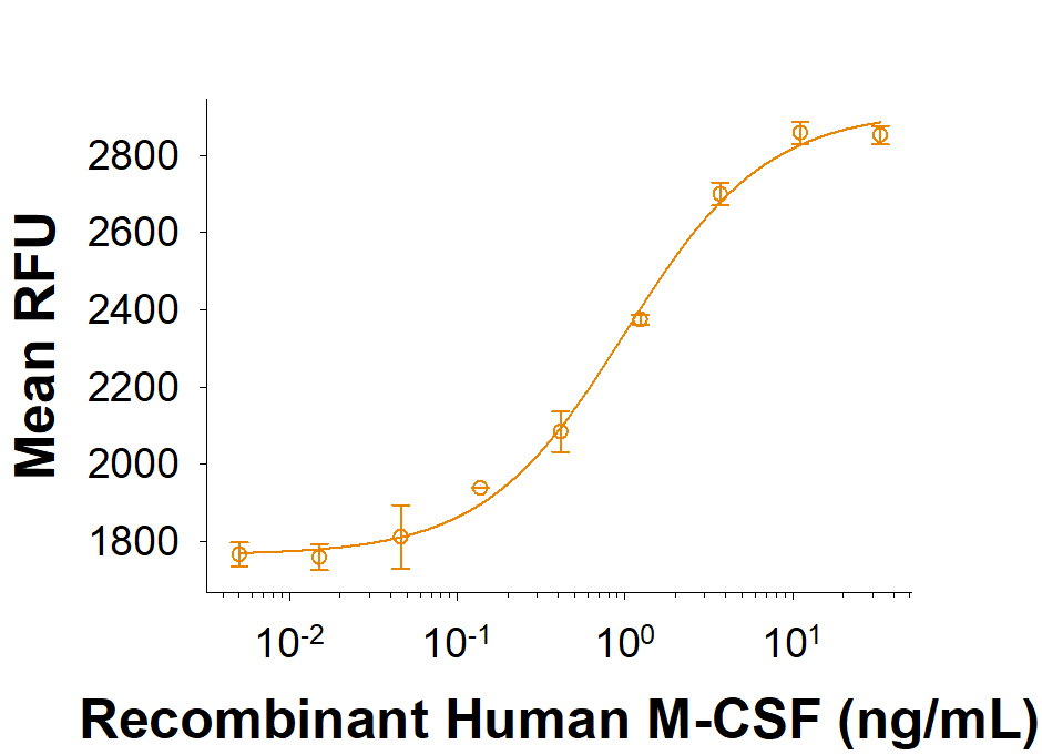
Measured in a cell proliferation assay using M‑NFS‑60 mouse myelogenous leukemia lymphoblast cells. The ED50 for this effect is 0.6-2.4 ng/mL.
Reconstitution Calculator
Background: M-CSF
M-CSF, also known as CSF-1, is a four alpha -helical-bundle cytokine that is the primary regulator of macrophage survival, proliferation and differentiation (1-3). M-CSF is also essential for the survival and proliferation of osteoclast progenitors (1, 4). M-CSF also primes and enhances macrophage killing of tumor cells and microorganisms, regulates the release of cytokines and other inflammatory modulators from macrophages, and stimulates pinocytosis (2, 3). M-CSF increases during pregnancy support implantation and growth of the decidua and placenta (5). Sources of M-CSF include fibroblasts, activated macrophages, endometrial secretory epithelium, bone marrow stromal cells and activated endothelial cells (1-5). The M-CSF receptor (c-fms) transduces its pleotropic effects and mediates its endocytosis. M-CSF mRNAs of various sizes occur (3-9). Full length human M-CSF transcripts encode a 522 amino acid (aa) type I transmembrane (TM) protein with a 464 aa extracellular region, a 21 aa TM domain, and a 37 aa cytoplasmic tail that forms a 140 kDa covalent dimer. Differential processing produces two proteolytically cleaved, secreted dimers. One is an N- and O-glycosylated 86 kDa dimer, while the other is modified by both glycosylation and chondroitin-sulfate proteoglycan (PG) to generate a 200 kDa subunit. Although PG-modified M-CSF can circulate, it may be immobilized by attachment to type V collagen (8). Shorter transcripts encode
M-CSF that lacks cleavage and PG sites and produces an N-glycosylated 68 kDa TM dimer and a slowly produced 44 kDa secreted dimer (7). Although forms may vary in activity and half-life, all contain the N-terminal 150 aa portion that is necessary and sufficient for interaction with the M-CSF receptor (10, 11). The first 223 aa of mature human M-CSF shares 88%, 86%, 81% and 74% aa identity with corresponding regions of canine, bovine, mouse and rat M-CSF, respectively (12, 13). Human M-CSF is active in the mouse, but mouse M-CSF is reported to be species specific.
- Pixley, F.J. and E.R. Stanley (2004) Trends Cell Biol. 14:628.
- Chitu, V. and E.R. Stanley (2006) Curr. Opin. Immunol. 18:39.
- Fixe, P. and V. Praloran (1997) Eur. Cytokine Netw. 8:125.
- Ryan, G.R. et al. (2001) Blood 98:74.
- Makrigiannakis, A. et al. (2006) Trends Endocrinol. Metab. 17:178.
- Nandi, S. et al. (2006) Blood 107:786.
- Rettenmier, C.W. and M.F. Roussel (1988) Mol. Cell Biol. 8:5026.
- Suzu, S. et al. (1992) J. Biol. Chem. 267:16812.
- Manos, M.M. (1988) Mol. Cell. Biol. 8:5035.
- Koths, K. (1997) Mol. Reprod. Dev. 46:31.
- Jang, M-H. et al. (2006) J. Immunol. 177:4055.
- Kawasaki, E.S. et al. (1985) Science 230: 291.
- Wong, G.G. et al. (1987) Science 235:1504.
Citations for Recombinant Human M-CSF (CHO-expressed) Protein
R&D Systems personnel manually curate a database that contains references using R&D Systems products. The data collected includes not only links to publications in PubMed, but also provides information about sample types, species, and experimental conditions.
3
Citations: Showing 1 - 3
Filter your results:
Filter by:
-
Acute and late administration of colony stimulating factor 1 attenuates chronic cognitive impairment following mild traumatic brain injury in mice
Authors: L Li, L Yerra, B Chang, V Mathur, A Nguyen, J Luo
Brain, Behavior, and Immunity, 2021-02-02;0(0):.
Species: Mouse
Sample Types: In Vivo
Applications: Bioassay -
Sequential conditioning-stimulation reveals distinct gene- and stimulus-specific effects of Type I and II IFN on human macrophage functions
Authors: Q Cheng, F Behzadi, S Sen, S Ohta, R Spreafico, R Teles, RL Modlin, A Hoffmann
Sci Rep, 2019-03-27;9(1):5288.
Species: Human
Sample Types: Whole Cells
Applications: Bioassay -
Lysosomal Protein Lamtor1 Controls Innate Immune Responses via Nuclear Translocation of Transcription Factor EB
Authors: Y Hayama, T Kimura, Y Takeda, S Nada, S Koyama, H Takamatsu, S Kang, D Ito, Y Maeda, M Nishide, S Nojima, H Sarashina-, T Hosokawa, Y Kinehara, Y Kato, T Nakatani, Y Nakanishi, T Tsuda, T Koba, M Okada, A Kumanogoh
J. Immunol., 2018-04-23;0(0):.
Species: Mouse
Sample Types: Whole Cells
Applications: Bioassay
FAQs
-
How long will recombinant human M-CSF last in cell culture?
End-users will need to determine the appropriate concentration and timing when adding Recombinant Human M-CSF Proteins to cell culture experiments. The addition of protein may be dependent on certain culture conditions, including the cell number, density, and media content. For techniques and methodologies, we recommend reviewing our list of publications under the Citations tab on the product-specific web page to find reported use of our products in similar experimental layouts.
-
Does recombinant human M-CSF show activity in mouse cells?
We evaluate the bioactivity of Recombinant Human M-CSF Protein (Catalog # 216-MCC) in a cell proliferation assay using mouse myelogenous leukemia lymphoblast cells. Catalog # 216-MCC exhibits an ED50 in the range of 0.6-2.4 ng/mL in this assay using mouse cells. We also offer the Recombinant Mouse M-CSF Protein (Catalog # 416-ML) for use in mouse assay systems.
Reviews for Recombinant Human M-CSF (CHO-expressed) Protein
There are currently no reviews for this product. Be the first to review Recombinant Human M-CSF (CHO-expressed) Protein and earn rewards!
Have you used Recombinant Human M-CSF (CHO-expressed) Protein?
Submit a review and receive an Amazon gift card.
$25/€18/£15/$25CAN/¥75 Yuan/¥2500 Yen for a review with an image
$10/€7/£6/$10 CAD/¥70 Yuan/¥1110 Yen for a review without an image


