SARS-CoV-2 NSP1 Antibody Summary
Met1-Gly180
Accession # YP_009725297.1
Scientific Data
 View Larger
NSP1 in HEK293 Human Cell Line Transfected with SARS-CoV-2. NSP1 was detected in immersion fixed HEK293 human embryonic kidney cell line transfected with SARS-CoV-2 (positive staining) and HEK293 human embryonic kidney cell line (non-transfected, negative staining) using Mouse Anti-SARS-CoV-2 NSP1 Monoclonal Antibody (Catalog # MAB10980) at 8 µg/mL for 3 hours at room temperature. Cells were stained using the NorthernLights™ 557-conjugated Anti-Mouse IgG Secondary Antibody (red; Catalog # NL007) and counterstained with DAPI (blue). Specific staining was localized to cytoplasm. View our protocol for Fluorescent ICC Staining of Cells on Coverslips.
 View Larger
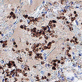
NSP1 in SARS-CoV-2 infected human lung. NSP1 was detected in immersion fixed paraffin-embedded sections of SARS-CoV-2 infected human lung using Mouse Anti-SARS-CoV-2 NSP1 Monoclonal Antibody (Catalog # MAB10980) at 5 µg/mL for 1 hour at room temperature followed by incubation with the Anti-Mouse IgG VisUCyte™ HRP Polymer Antibody (Catalog # VC001). Before incubation with the primary antibody, tissue was subjected to heat-induced epitope retrieval using Antigen Retrieval Reagent-Basic (Catalog # CTS013). Tissue was stained using DAB (brown) and counterstained with hematoxylin (blue). Specific staining was localized to immunoreactive profiles scattered throughout the tissue. View our protocol for IHC Staining with VisUCyte HRP Polymer Detection Reagents.
 View Larger
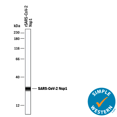
Detection of SARS-CoV-2 NSP1 by Simple WesternTM. Simple Western lane view shows recombinant SARS-CoV-2 Nsp1, loaded at 0.2 mg/mL. A specific band was detected for NSP1 at approximately 28 kDa (as indicated) using 20 µg/mL of Mouse Anti-SARS-CoV-2 NSP1 Monoclonal Antibody (Catalog # MAB10980). This experiment was conducted under reducing conditions and using the 12-230 kDa separation system.
 View Larger
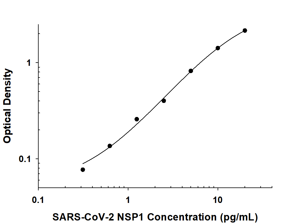
SARS-CoV-2 NSP1 ELISA Standard Curve. Recombinant SARS-CoV-2 NSP1 protein was serially diluted 2-fold and captured by Mouse Anti-SARS-CoV-2 NSP1 Monoclonal Antibody (Catalog # MAB10980) coated on a Clear Polystyrene Microplate (Catalog # DY990). Mouse Anti-SARS-CoV-2 NSP1 Monoclonal Antibody (Catalog # MAB10991) was biotinylated and incubated with the protein captured on the plate. Detection of the standard curve was achieved by incubating Streptavidin-HRP (Catalog # DY998) followed by Substrate Solution (Catalog # DY999) and stopping the enzymatic reaction with Stop Solution (Catalog # DY994).
Reconstitution Calculator
Preparation and Storage
- 12 months from date of receipt, -20 to -70 °C as supplied.
- 1 month, 2 to 8 °C under sterile conditions after reconstitution.
- 6 months, -20 to -70 °C under sterile conditions after reconstitution.
Background: NSP1
Non-structural protein 1 (NSP1) is one of several functional proteins released by ORF1a-encoded protease cleavage of the pp1a and pp1ab replicase polyproteins expressed from the coronavirus (CoV) genome (1). The NSPs are involved in the replication and transcription of the viral RNA and not incorporated within the virion particles. Coronaviruses include various highly pathogenic strains such as SARS-CoV, MERS-CoV and SARS-CoV2 that have had significant impact on humans as well as strains that have negatively impacted livestock. NSP1, also known as the host shutoff factor, is a small 180 amino acid highly conserved protein among the first to be expressed following cell entry. It is composed of an N-terminal domain, a linker region, and a C-terminal domain with a conserved KH motif that is required and sufficient for specific contacts with ribosomes (2, 3). The C-terminal domain binds tightly to and sterically occludes the entrance region of the mRNA channel in free 40S subunits, 43S pre-initiation complex, and empty, non-translating 80S ribosomes (2, 3) to inhibit translation. NSP1 blocks the innate immune response through suppression of host gene expression. By preventing translation of interferon and downstream signaling responses, it plays a key role in immune evasion (2, 4, 5). NSP1 also induces endonucleolytic cleavage of host mRNAs (6,7). Concomitant translation of more efficiently recognized untranslated regions (UTR) of viral mRNA along with resistance to endonucleolytic cleavage (7) is thought to lead to a switch to production of viral mRNA over host cell mRNA during an infection (3,8). Given the critical role NSP1 plays in virulence, it is an attractive target for small-molecule inhibition and vaccination development (9, 10).
- Snijder, E.J. et al. (2016) Adv. Virus Res 96:59.
- Thoms, M. et al. (2020) Science 369:1249.
- Schubert, K. et al. (2020) Nat. Struct. Mol. Biol. 27:959.
- Narayanan, K. et al. (2008) J. Virol. 82:4471.
- Jauregui, A.R. et al. (2013) PLoS One. 8:e62416.
- Kamitani, W. et al. (2006) Proc. Natl. Acad. Sci. USA 103:12885.
- Huang, C. et al. (2011) PLoS Pathog. 7:e1002433.
- Hartenian, E. et al. (2020) J. Biol. Chem. 295:12910.
- Zust, R. et al. (2007) PLoS Pathog. 3:e109.
- De Lima Menezes, G. and R.A. da Silva (2020) J. Biomol. Struct. Dyn. (In press).
Product Datasheets
FAQs
No product specific FAQs exist for this product, however you may
View all Antibody FAQsReviews for SARS-CoV-2 NSP1 Antibody
There are currently no reviews for this product. Be the first to review SARS-CoV-2 NSP1 Antibody and earn rewards!
Have you used SARS-CoV-2 NSP1 Antibody?
Submit a review and receive an Amazon gift card.
$25/€18/£15/$25CAN/¥75 Yuan/¥2500 Yen for a review with an image
$10/€7/£6/$10 CAD/¥70 Yuan/¥1110 Yen for a review without an image

