VisULite ECL Western Blotting Substrate
VisULite ECL Western Blotting Substrate Summary
Kit Summary
Ready-to-use high sensitivity Enhanced Chemiluminescent (ECL) substrate.
Stability and Storage
Store in the dark at 2 °C to 8 °C. DO NOT FREEZE.
Specifications
Product Datasheets
Scientific Data
 View Larger
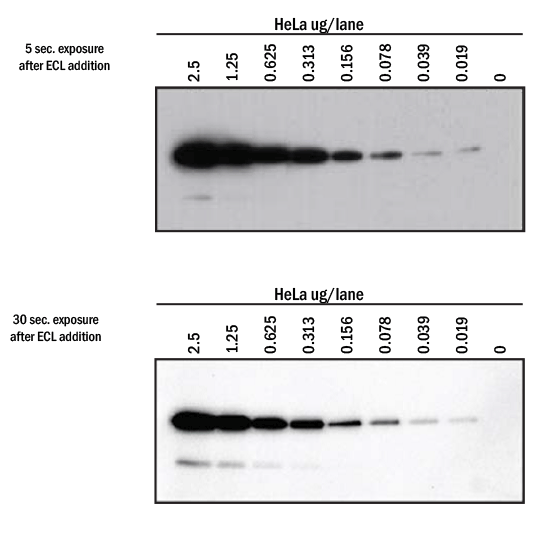
Detection of GAPDH in HeLa cell lysates visualized with VisULiteTM ECL Western Blotting Substrate using X-ray film or CCD imaging system. A two-fold dilution series of HeLa whole cell lysate was prepared and loaded at 2.5, 1.25, 0.625, 0.313, 0.156, 0.078, 0.039 and 0.019 µg per lane. Samples were transferred onto PVDF membrane and probed with 0.1 µg/mL of Mouse Anti-Human/Mouse/Rat GAPDH Monoclonal Antibody (R&D Systems®, Catalog # MAB5718) followed by HRP-conjugated Anti-Mouse IgG Secondary Antibody (R&D Systems®, Catalog # HAF018). VisULite™ ECL substrate was applied for 1 minute, followed by exposure to a standard sensitivity film (left) or image capture using ProteinSimple® FluorChem™ M (right). Lanes are visualized on a 5 second exposure (film) or 30 second exposure (imager).
 View Larger
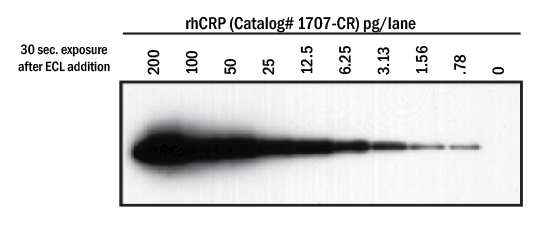
Detection of Recombinant Human CRP visualized with VisULite™ ECL Western Blotting Substrate. A two-fold dilution series was prepared using rhCRP (R&D Systems®, Catalog # 1707-CR) and loaded at 200, 100, 50, 25, 12.5, 6.25, 3.13, 1.56 and 0.78 pg per lane. Samples were transferred onto PVDF membrane and probed with 0.5 µg/mL of Sheep Anti-Human CRP (R&D Systems®, Catalog # AF1707) followed by HRP-conjugated Anti-Sheep IgG Secondary Antibody (R&D Systems®, Catalog # HAF016). VisULite™ ECL substrate was applied for 1 minute, followed by exposure to a standard sensitivity film for 30 seconds.
 View Larger
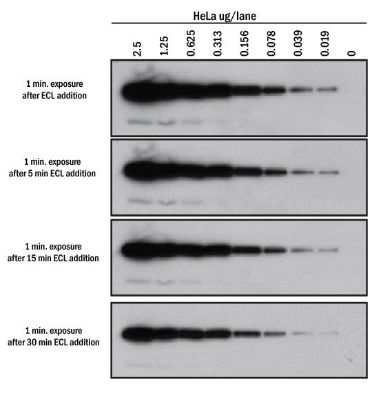
Signal duration with VisULite™ ECL Western Blotting Substrate. A two-fold dilution series of HeLa whole cell lysate was prepared, loaded, transferred and probed with Mouse Anti-Human/Mouse/Rat GAPDH Monoclonal Antibody (R&D Systems®, Catalog # MAB5718) as in Figure 1 (above). VisULiteTM ECL substrate was applied for 1 minute to the PVDF membrane, followed by exposure to a standard sensitivity film for 1 minute at the indicated timepoints. All lanes are visualized on a 1 minute exposure 0 to 30 minutes after VisULite™ ECL substrate addition to the PVDF membrane.
Assay Procedure
Refer to the product datasheet for complete product details.
Precaution
See MSDS.
Reagents Provided
200 mL VisULite™ ECL Western Blotting Substrate
Procedure Overview
Run SDS-PAGE gel and transfer to nitrocellulose or PVDF by established laboratory protocol.
Label and block the membrane.
Incubate the membrane with primary antibody. Note: High sensitivity of substrate may require adjusting established parameters for primary and secondary antibody.
Wash and incubate with appropriate HRP-labeled secondary antibody.
Wash and, at the completion of the wash, drain off all liquid and gently blot dry on a lab wipe or filter paper. Note: Do not let membrane dry completely.
Place membrane on clean plastic wrap and add approximately 100 µL of VisULite™ ECL Western Blotting Substrate per square centimeter of membrane. Allow to incubate for 1 minute.
Drain excess substrate and gently blot dry as above.
Place membrane in CCD camera system and expose according to manufacturer’s instructions or place membrane on clean plastic wrap (or equivalent) and expose to film.
Adjust exposure times as needed.
Limitations
- FOR LABORATORY RESEARCH USE ONLY. NOT FOR USE IN DIAGNOSTIC PROCEDURES.
- This reagent should not be used beyond the expiration date indicated on the label.
- Results may vary due to variations in blotting conditions.
- Note: Because VisULite™ ECL Western Blotting Substrate is a highly sensitive substrate, optimization of primary and secondary antibodies may be required to generate consistently reproducible results with strong signals and low backgrounds. See www.rndsystems.com/resources/protocols/western-blot-cell-lysate-protocol for additional optimization tips and hints that may further improve performance.
- Do not allow membrane to completely dry during procedure.
- Do not use Sodium Azide in any buffers used for detection. Azide is an inhibitor of HRP.
- Work quickly once the membrane has been exposed to VisULite™ ECL Western Blotting Substrate to capture maximum signal.
Citations for VisULite ECL Western Blotting Substrate
R&D Systems personnel manually curate a database that contains references using R&D Systems products. The data collected includes not only links to publications in PubMed, but also provides information about sample types, species, and experimental conditions.
5
Citations: Showing 1 - 5
Filter your results:
Filter by:
-
Gao-Zi-Yao improves learning and memory function in old spontaneous hypertensive rats
Authors: MX Han, WY Jiang, Y Jiang, LH Wang, R Xue, GX Zhang, JW Chen
BMC complementary medicine and therapies, 2022-05-28;22(1):147. 2022-05-28
-
Effects of Tiaozhi Granule on Regulation of Autophagy Levels in HUVECs
Authors: YY Wu, C Chen, X Yu, XD Zhao, RQ Bao, JY Yu, GX Zhang, JW Chen
Evid Based Complement Alternat Med, 2018-07-12;2018(0):1765731. 2018-07-12
-
Variations in the Peritrophic Matrix Composition of Heparan Sulphate from the Tsetse Fly, Glossina morsitans morsitans
Authors: E Rogerson, J Pelletier, A Acosta-Ser, C Rose, S Taylor, S Guimond, M Lima, M Skidmore, E Yates
Pathogens, 2018-03-19;7(1):. 2018-03-19
-
Tanshinone IIA Sodium Sulfonate Attenuates LPS-Induced Intestinal Injury in Mice
Authors: XJ Yang, JX Qian, Y Wei, Q Guo, J Jin, X Sun, SL Liu, CF Xu, GX Zhang
Gastroenterol Res Pract, 2018-03-08;2018(0):9867150. 2018-03-08
-
The Development of an Angiogenic Protein Signature in Ovarian Cancer Ascites as a Tool for Biologic and Prognostic Profiling
Authors: Sofia-Paraskevi Trachana
PLoS ONE, 2016-06-03;11(6):e0156403. 2016-06-03
FAQs
No product specific FAQs exist for this product, however you may
View all Supplemental Reagent FAQsReviews for VisULite ECL Western Blotting Substrate
There are currently no reviews for this product. Be the first to review VisULite ECL Western Blotting Substrate and earn rewards!
Have you used VisULite ECL Western Blotting Substrate?
Submit a review and receive an Amazon gift card.
$25/€18/£15/$25CAN/¥75 Yuan/¥2500 Yen for a review with an image
$10/€7/£6/$10 CAD/¥70 Yuan/¥1110 Yen for a review without an image
