Human IGFBP-rp1/IGFBP-7 Biotinylated Antibody Summary
Arg98-Arg277
Accession # AAA16187
Applications
Please Note: Optimal dilutions should be determined by each laboratory for each application. General Protocols are available in the Technical Information section on our website.
Scientific Data
 View Larger
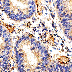
IGFBP-rp1/IGFBP-7 in Human Colon Cancer Tissue. IGFBP-rp1/IGFBP-7 was detected in immersion fixed paraffin-embedded sections of human colon cancer tissue using Goat Anti-Human IGFBP-rp1/IGFBP-7 Biotinylated Antigen Affinity-purified Polyclonal Antibody (Catalog # BAF1334) at 15 µg/mL overnight at 4 °C. Before incubation with the primary antibody, tissue was subjected to heat-induced epitope retrieval using Antigen Retrieval Reagent-Basic (Catalog # CTS013). Tissue was stained using the Anti-Goat HRP-DAB Cell & Tissue Staining Kit (brown; Catalog # CTS008) and counterstained with hematoxylin (blue). Specific staining was localized to endothelial cells. View our protocol for Chromogenic IHC Staining of Paraffin-embedded Tissue Sections.
Reconstitution Calculator
Preparation and Storage
- 12 months from date of receipt, -20 to -70 °C as supplied.
- 1 month, 2 to 8 °C under sterile conditions after reconstitution.
- 6 months, -20 to -70 °C under sterile conditions after reconstitution.
Background: IGFBP-rp1/IGFBP-7
IGFBP-rp1, also known as Mac25/Angiomodulin (AGM), tumor-derived adhesion factor (TAF) and prostacyclin-stimulating factor (PSF), is a secreted protein that contains three protein domain modules. Human IGFBP-rp1 cDNA encodes 282 amino acid (aa) residue precursor protein with a putative 26 aa signal peptide. Mature IGFBP-rp1 is a glycosylated protein with an N-terminal IGFBP domain, followed by a Kazal-type serine proteinase inhibitor domain and a C-terminal immunoglobulin‑like C2-type domain. The similarity of IGFBP-rp1 with the IGFBPs is confined to the N-terminal IGFBP domain, which contains all 12 of the conserved cysteine residues found in IGFBP1 through 5. Human and mouse IGFBP-rP1 are highly homologous. Discounting a segment of 43 aa near the N-terminus that is missing in the mouse homologue, human and mouse IGFBP-rp1 share 94% aa sequence identity. IGFBP-rp1 is expressed in many normal tissues and in cancer cells. It is abundantly expressed in high endothelial venules (HEVs) of blood vessels in the secondary lymphoid tissues. The expression of IGFBP-rp1 is up‑regulated in senescing epithelial cells and by retinoic acid. IGFBP-rp1 binds IGF and insulin with very low affinity and has been shown to enhance the mitogenic actions of IGF and insulin. IGFBP-rp1 also has IGF/insulin-independent activities. It interacts with heparan sulfate proteoglycans, type IV collagen, and specific chemokines. IGFBP‑rp1 supports weak cell adhesion, promotes cell spreading on type IV collagen, and stimulates the production of the potent vasodilator PGI2.. It modulates tumor cell growth and has also been implicated in angiogenesis. IGFBP-rp1 is proteolytically cleaved between lysine 97 and alanine 98. Cleaved IGFBP-rp1 has enhanced cell attachment activity but can no longer bind IGF/insulin (1‑3).
- Hwa, V. et al. (1999) Endocrinology Rev. 20:761.
- Nagakubo, D. et al. (2003) J. Immunol. 171:553.
- Ahmed, S. et al. (2003) Biochem. Biophys. Res. Commun. 310:612.
Product Datasheets
FAQs
No product specific FAQs exist for this product, however you may
View all Antibody FAQsReviews for Human IGFBP-rp1/IGFBP-7 Biotinylated Antibody
There are currently no reviews for this product. Be the first to review Human IGFBP-rp1/IGFBP-7 Biotinylated Antibody and earn rewards!
Have you used Human IGFBP-rp1/IGFBP-7 Biotinylated Antibody?
Submit a review and receive an Amazon gift card.
$25/€18/£15/$25CAN/¥75 Yuan/¥2500 Yen for a review with an image
$10/€7/£6/$10 CAD/¥70 Yuan/¥1110 Yen for a review without an image

