Mouse Semaphorin 7A Antibody Summary
Gln45-Ala646
Accession # Q9QUR8
Applications
Please Note: Optimal dilutions should be determined by each laboratory for each application. General Protocols are available in the Technical Information section on our website.
Scientific Data
 View Larger
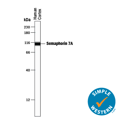
Detection of Human Semaphorin 7A by Simple WesternTM. Simple Western lane view shows lysates of human brain (cortex) tissue, loaded at 0.2 mg/mL. A specific band was detected for Semaphorin 7A at approximately 109 kDa (as indicated) using 2.5 µg/mL of Goat Anti-Mouse Semaphorin 7A Antigen Affinity-purified Polyclonal Antibody (Catalog # AF1835) followed by 1:50 dilution of HRP-conjugated Anti-Goat IgG Secondary Antibody (Catalog # HAF109). This experiment was conducted under reducing conditions and using the 12-230 kDa separation system.
 View Larger
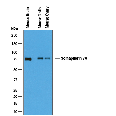
Detection of Mouse Semaphorin 7A by Western Blot. Western blot shows lysates of mouse brain tissue, mouse testis tissue, and mouse ovary tissue. PVDF membrane was probed with 0.25 µg/mL of Goat Anti-Mouse Semaphorin 7A Antigen Affinity-purified Polyclonal Antibody (Catalog # AF1835) followed by HRP-conjugated Anti-Goat IgG Secondary Antibody (Catalog # HAF019). A specific band was detected for Semaphorin 7A at approximately 75 kDa (as indicated). This experiment was conducted under reducing conditions and using Immunoblot Buffer Group 1.
Reconstitution Calculator
Preparation and Storage
- 12 months from date of receipt, -20 to -70 °C as supplied.
- 1 month, 2 to 8 °C under sterile conditions after reconstitution.
- 6 months, -20 to -70 °C under sterile conditions after reconstitution.
Background: Semaphorin 7A
Semaphorin 7A (Sema7A, designated CD108, previously Sema K1 or Sema L), is an ~80 kDa membrane-anchored glycoprotein that is a member of the Semaphorin family of axon guidance molecules (1 - 4). On human erythrocytes, it is the John Milton Hagen (JMH) blood group antigen (4). Sema7A is the only known Class 7 or glycophosphatidylinositol (GPI)-linked semaphorin; its expression is concentrated in the brain, spleen and thymus (1 - 5). Mouse Sema7A cDNA encodes a 44 amino acid (aa) signal sequence, a 602 aa extracellular domain (ECD) including Sema and C2-type Ig-like domains, and an 18 aa propeptide/GPI membrane anchor signal sequence. Mature mouse Sema7A shares 89%, 98%, 85%, 86% and 89% aa identity with corresponding human, rat, bovine, canine and equine Sema7A, respectively. The Sema7A sema domain contains an RGD integrin interaction motif (4). Although it binds plexin-C1 in vitro and may be coexpressed with it, many of its activities depend on interaction with beta 1 integrins such as alpha 1 beta 1 (6 - 10). Sema7A signaling through the two receptors may cause opposing effects (8). Sema7A is an immune semaphorin, with expression and activity on CD4+CD8+ thymocytes, activated T cells, macrophages and microglia (2, 9 - 12). T cell Sema7A interacts with monocytic cells, stimulating their chemotaxis, production of pro-inflammatory cytokines, and dendritic differentiation (5, 6). However, on the T cells themselves, Sema7A downregulates TCR signaling by promoting TCR internalization, modulating T cell responses (9). In lung macrophages, Sema7A is induced by TGF-beta and participates in TGF-beta -induced lung fibrosis (12). Sema7A is also expressed on pre-osteoblasts and osteoclasts, where it promotes migration and fusion, respectively; on keratinocytes, where it promotes melanocyte spreading and dendricity; and on some neurons, for example, promoting axon outgrowth in the developing olfactory tract (8, 10, 13).
- Yazdani, U. and J.R. Terman (2006) Genome Biol. 7:211.
- Kikutani, H. et al. (2007) Adv. Immunol. 93:121.
- Sato, Y. and (1998) Biochim. Biophys. Acta 1443:419.
- Yamada, A. et al. (1999) J. Immunol. 162:4094.
- Holmes, S. et al. (2002) Scand. J. Immunol. 56:270.
- Suzuki, K. et al. (2007) Nature 446:680.
- Pasterkamp, R.J. et al. (2007) BMC Dev. Biol. 7:98.
- Scott, G.A. et al. (2007) J. Invest. Dermatol. 128:151.
- Czopik, A.K. et al. (2006) Immunity 24:591.
- Pasterkamp, R.J. et al. (2003) Nature 424:398.
- Mine, T. et al. (2000) Tissue Antigens 55:429.
- Kang, H-R. et al. (2007) J. Exp. Med. 204:1083.
- Delorme, G. et al. (2005) Biol. Cell 97:589.
Product Datasheets
Citations for Mouse Semaphorin 7A Antibody
R&D Systems personnel manually curate a database that contains references using R&D Systems products. The data collected includes not only links to publications in PubMed, but also provides information about sample types, species, and experimental conditions.
3
Citations: Showing 1 - 3
Filter your results:
Filter by:
-
Sema7A/PlxnCl signaling triggers activity-dependent olfactory synapse formation
Authors: N Inoue, H Nishizumi, H Naritsuka, H Kiyonari, H Sakano
Nat Commun, 2018-05-09;9(1):1842.
Species: Mouse
Sample Types: Whole Tissue
Applications: IHC -
Rewiring the taste system
Authors: H Lee, LJ Macpherson, CA Parada, CS Zuker, NJP Ryba
Nature, 2017-08-09;548(7667):330-333.
Species: Mouse
Sample Types: Whole Tissue
Applications: IHC -
Semaphorin7A regulates neuroglial plasticity in the adult hypothalamic median eminence.
Authors: Parkash J, Messina A, Langlet F, Cimino I, Loyens A, Mazur D, Gallet S, Balland E, Malone S, Pralong F, Cagnoni G, Schellino R, De Marchis S, Mazzone M, Pasterkamp R, Tamagnone L, Prevot V, Giacobini P
Nat Commun, 2015-02-27;6(0):6385.
Species: Mouse
Sample Types: Tissue Homogenates, Whole Tissue
Applications: IHC, Western Blot
FAQs
No product specific FAQs exist for this product, however you may
View all Antibody FAQsReviews for Mouse Semaphorin 7A Antibody
There are currently no reviews for this product. Be the first to review Mouse Semaphorin 7A Antibody and earn rewards!
Have you used Mouse Semaphorin 7A Antibody?
Submit a review and receive an Amazon gift card.
$25/€18/£15/$25CAN/¥75 Yuan/¥2500 Yen for a review with an image
$10/€7/£6/$10 CAD/¥70 Yuan/¥1110 Yen for a review without an image

