Human/Mouse COX4-I1 Antibody Summary
Ala23-Lys169
Accession # P13073
Applications
Please Note: Optimal dilutions should be determined by each laboratory for each application. General Protocols are available in the Technical Information section on our website.
Scientific Data
 View Larger
Detection of Human/Mouse COX4-I1 by Western Blot. Western blot shows lysates of human brain tissue, Jurkat human acute T cell leukemia cell line, HepG2 human hepatocellular carcinoma cell line, and BaF3 mouse pro-B cell line. PVDF membrane was probed with 1 µg/mL of Goat Anti-Human/Mouse COX4-I1 Antigen Affinity-purified Polyclonal Antibody (Catalog # AF5814) followed by HRP-conjugated Anti-Goat IgG Secondary Antibody (Catalog # HAF109). A specific band was detected for COX4-I1 at approximately 18 kDa (as indicated). This experiment was conducted under reducing conditions and using Immunoblot Buffer Group 2.
 View Larger
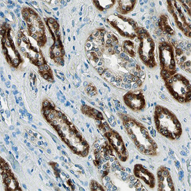
COX4-I1 in Human Kidney. COX4-I1 was detected in immersion fixed paraffin-embedded sections of normal human kidney using Goat Anti-Human/Mouse COX4-I1 Antigen Affinity-purified Polyclonal Antibody (Catalog # AF5814) at 10 µg/mL overnight at 4 °C. Before incubation with the primary antibody, tissue was subjected to heat-induced epitope retrieval using Antigen Retrieval Reagent-Basic (Catalog # CTS013). Tissue was stained using the Anti-Goat HRP-DAB Cell & Tissue Staining Kit (brown; Catalog # CTS008) and counterstained with hematoxylin (blue). View our protocol for Chromogenic IHC Staining of Paraffin-embedded Tissue Sections.
 View Larger
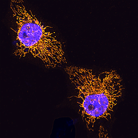
COX4‑I1 in HeLa Human Cell Line. COX4-I1 was detected in immersion fixed HeLa human cervical epithelial carcinoma cell line using Goat Anti-Human/Mouse COX4-I1 Antigen Affinity-purified Polyclonal Antibody (Catalog # AF5814) at 5 µg/mL for 3 hours at room temperature. Cells were stained using the NorthernLights™ 557-conjugated Anti-Goat IgG Secondary Antibody (yellow; Catalog # NL001) and counterstained with DAPI (blue). Specific staining was localized to mitochondria. View our protocol for Fluorescent ICC Staining of Cells on Coverslips.
 View Larger
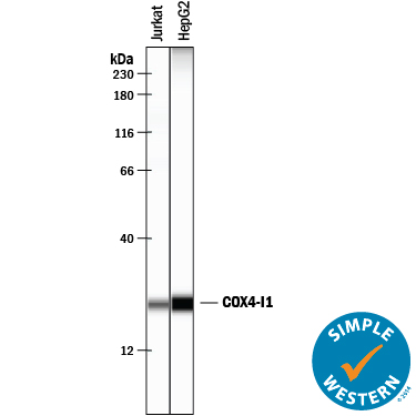
Detection of Human COX4‑I1 by Simple WesternTM. Simple Western lane view shows lysates of Jurkat human acute T cell leukemia cell line and HepG2 human hepatocellular carcinoma cell line, loaded at 0.2 mg/mL. A specific band was detected for COX4-I1 at approximately 24 kDa (as indicated) using 10 µg/mL of Goat Anti-Human/Mouse COX4-I1 Antigen Affinity-purified Polyclonal Antibody (Catalog # AF5814) followed by 1:50 dilution of HRP-conjugated Anti-Goat IgG Secondary Antibody (Catalog # HAF109). This experiment was conducted under reducing conditions and using the 12-230 kDa separation system.
Reconstitution Calculator
Preparation and Storage
- 12 months from date of receipt, -20 to -70 °C as supplied.
- 1 month, 2 to 8 °C under sterile conditions after reconstitution.
- 6 months, -20 to -70 °C under sterile conditions after reconstitution.
Background: COX4-I1
Cytochrome c oxidase subunit 4 isoform 1 (COX-4I1) is a 21‑22 kDa member of the cytochrome c oxidase IV family of proteins. It is a component of COX, an inner mitochondrial membrane multimeric dimer that catalyzes the transfer of electrons from cytochrome c to dioxygen. COX-4I1 is the largest of 10 distinct nuclear DNA‑encoded subunits that form each COX monomer. Human COX4-I1 is 169 amino acids (aa) in length and possesses a mitochondrial transit peptide between aa 1‑22, an ATP binding site (aa 42; 95‑100), and multiple subunit interface sequences. The ATP binding site is suggested to make COX-4I1 a regulatory subunit within the COX complex. There are two potential splice variants for COX-4I1 that show a three aa substitution for aa 81‑169, and a single Gly substitution for aa 125‑169. COX‑4I2, the product of a different gene, shares only 50% aa identity with COX-4I1. Over amino acid 23‑169, human COX4‑1 shares 78% aa identity with mouse COX‑4I1.
Product Datasheets
Citation for Human/Mouse COX4-I1 Antibody
R&D Systems personnel manually curate a database that contains references using R&D Systems products. The data collected includes not only links to publications in PubMed, but also provides information about sample types, species, and experimental conditions.
1 Citation: Showing 1 - 1
-
Sarcolipin Signaling Promotes Mitochondrial Biogenesis and Oxidative Metabolism in Skeletal Muscle
Authors: SK Maurya, JL Herrera, SK Sahoo, FCG Reis, RB Vega, DP Kelly, M Periasamy
Cell Rep, 2018-09-11;24(11):2919-2931.
Species: Mouse
Sample Types: Whole Tissue
Applications: IHC
FAQs
No product specific FAQs exist for this product, however you may
View all Antibody FAQsReviews for Human/Mouse COX4-I1 Antibody
Average Rating: 4 (Based on 1 Review)
Have you used Human/Mouse COX4-I1 Antibody?
Submit a review and receive an Amazon gift card.
$25/€18/£15/$25CAN/¥75 Yuan/¥2500 Yen for a review with an image
$10/€7/£6/$10 CAD/¥70 Yuan/¥1110 Yen for a review without an image
Filter by:
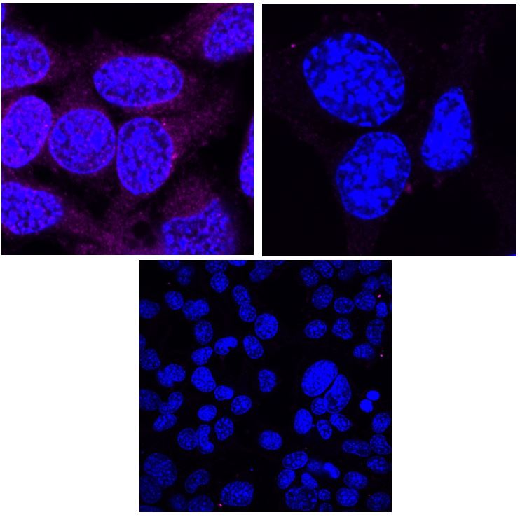
NSC-34 cells were cultured and fixed in methanol before immunofluorescence staining was conducted. COXIV antibody was used at 1 in 50 and no primary antibody and secondary antibody controls were conducted. Imaging was done on a confocal microscope.


