Human Neurofascin Antibody Summary
Ile25-Trp1039
Accession # NP_055905
Applications
Please Note: Optimal dilutions should be determined by each laboratory for each application. General Protocols are available in the Technical Information section on our website.
Scientific Data
 View Larger
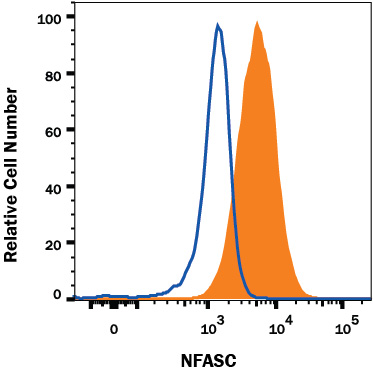
Detection of NFASC in U87-MG Human Cell Line by Flow Cytometry. U87-MG human cell line was stained with Mouse Anti-Human NFASC Monoclonal Antibody (Catalog # MAB8208, filled histogram) or isotype control antibody (Catalog # MAB002, open histogram), followed by PE-conjugated Anti-Mouse IgG Secondary Antibody (Catalog # F0102B). View our protocol for Staining Membrane-associated Proteins.
Reconstitution Calculator
Preparation and Storage
- 12 months from date of receipt, -20 to -70 °C as supplied.
- 1 month, 2 to 8 °C under sterile conditions after reconstitution.
- 6 months, -20 to -70 °C under sterile conditions after reconstitution.
Background: Neurofascin
Neurofascin, a type I transmembrane glycoprotein, is a member of the L1 family of cell adhesion molecules (CAMs) (1-4). L1CAM family members are composed of an extracellular domain (ECD) that contains six immunoglobulin (Ig)-like domains and multiple fibronectin type III repeats, followed by transmembrane and cytoplasmic domains (2, 4, 5). Multiple isoforms of Neurofascin, including NF155, NF166, NF180, and NF186, can be generated by alternative splicing with a predicted range of approximately 70 kDa to 150 kDa (4). These isoforms differ in the combination of fibronectin type III repeats, as well as in the presence of a proline-, alanine-, and threonine-rich segment (PAT domain) located just after the fourth fibronectin type III repeat (4). This recombinant human Neurofascin protein corresponds to rat isoform NF155 and shares 96% amino acid sequence identity with comparable regions of rat and mouse Neurofascin. In rats, NF155 is transiently expressed by oligodendrocytes and Schwann cells during axon myelination (6, 7). NF155 clusters in paranodal regions of oligodendroglia and binds to the Caspr-Contactin complex located on the adjacent axon to form and stabilize paranodal axoglial junctions (8-11). It has been suggested that the ECD of NF155 must be cleaved from oligodendroglia membranes to form and/or stabilize the paranodal structure (12). NF155 has also been shown to promote neuronal adhesion and neurite outgrowth in rats and chickens (12-14). Alterations in NF155 expression have been associated with multiple sclerosis (15).
- Rathjen, F.G. et al. (1987) Cell 51:841.
- Volkmer, H. et al. (1992) J. Cell Biol. 118:149.
- Sherman, D.L. and P.J. Brophy (2005) Nat. Rev. Neurosci. 6:683.
- Kriebel, M. et al. (2012) Int. J. Biochem. Cell Biol. 44:694.
- Liu, H. et al. (2011) J. Biol. Chem. 286:797.
- Collinson, J.M. et al. (1998) Glia 23:11.
- Basak, S. et al. (2007) Dev. Biol. 311:408.
- Charles, P. et al. (2002) Curr. Biol. 12:217.
- Schafer, D.P. et al. (2004) J. Neurosci. 24:3176.
- Maier, O. et al. (2005) Mol. Cell. Neurosci. 28:390.
- Sherman, D.L. et al. (2005) Neuron 48:737.
- Maier, O. et al. (2006) Exp. Cell Res. 312:500.
- Volkmer, H. et al. (1996) J. Cell Biol. 135:1059.
- Koticha, D. et al. (2005) Mol. Cell. Neurosci. 30:137.
- Howell, O.W. et al. (2006) Brain 129:3173.
Product Datasheets
FAQs
No product specific FAQs exist for this product, however you may
View all Antibody FAQsReviews for Human Neurofascin Antibody
There are currently no reviews for this product. Be the first to review Human Neurofascin Antibody and earn rewards!
Have you used Human Neurofascin Antibody?
Submit a review and receive an Amazon gift card.
$25/€18/£15/$25CAN/¥75 Yuan/¥2500 Yen for a review with an image
$10/€7/£6/$10 CAD/¥70 Yuan/¥1110 Yen for a review without an image

