Human PD-1 Biotinylated Antibody Summary
Leu25-Gln167
Accession # Q8IX89
Applications
Human PD-1 Sandwich Immunoassay
Please Note: Optimal dilutions should be determined by each laboratory for each application. General Protocols are available in the Technical Information section on our website.
Scientific Data
 View Larger
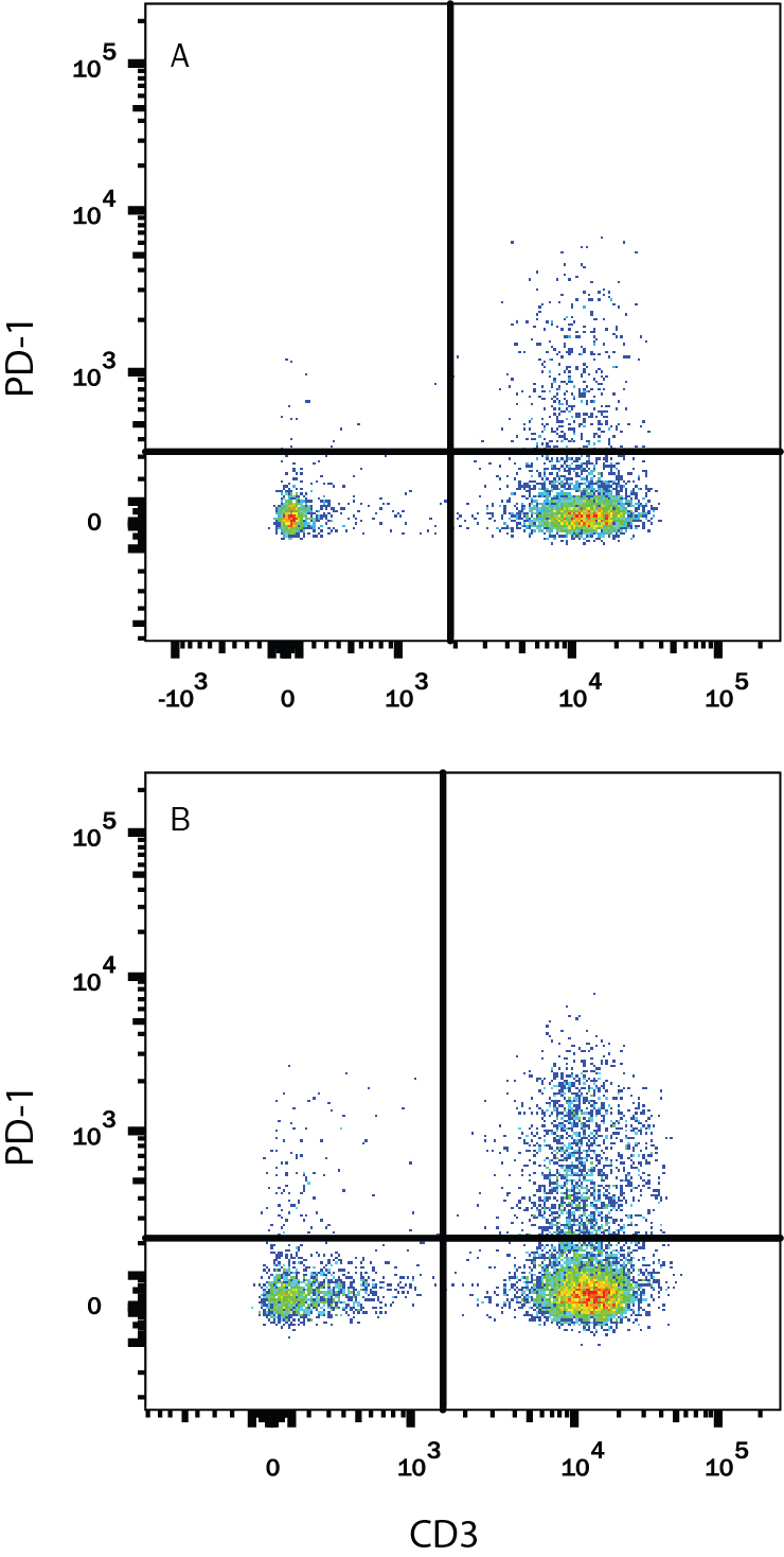
Detection of PD‑1 in Human PBMCs treated with PHA by Flow Cytometry. Human peripheral blood mononuclear cells (PBMCs) either (A) untreated or (B) treated with 5 µg/mL PHA overnight were stained with Goat Anti-Human PD-1 Biotinylated Antigen Affinity-purified Polyclonal Antibody (Catalog # BAF1086) followed by Streptavidin-Phycoerythrin (Catalog # F0040) and Mouse Anti-Human CD3e APC-conjugated Monoclonal Antibody (Catalog # FAB100A). Quadrant markers were set based on control antibody staining (Catalog # BAF108). View our protocol for Staining Membrane-associated Proteins.
Reconstitution Calculator
Preparation and Storage
- 12 months from date of receipt, -20 to -70 °C as supplied.
- 1 month, 2 to 8 °C under sterile conditions after reconstitution.
- 6 months, -20 to -70 °C under sterile conditions after reconstitution.
Background: PD-1
Programmed Death-1 (PD-1) is a type I transmembrane protein belonging to the CD28/CTLA-4 family of immunoreceptors that mediate signals for regulating immune responses (1). Members of the CD28/CTLA-4 family have been shown to either promote T cell activation (CD28 and ICOS) or downregulate T cell activation (CTLA-4 and PD-1) (2). PD-1 is expressed on activated T cells, B cells, myeloid cells, and on a subset of thymocytes. In vitro, ligation of PD-1 inhibits TCR-mediated T cell proliferation and production of IL-1, IL-4, IL-10, and IFN-gamma. In addition, PD-1 ligation also inhibits BCR mediated signaling. PD-1 deficient mice have a defect in peripheral tolerance and spontaneously develop autoimmune diseases (2, 3).
Two B7 family proteins, PD-L1 (also called B7-H1) and PD-L2 (also known as B7-DC), have been identified as PD-1 ligands. Unlike other B7 family proteins, both PD‑L1 and PD‑L2 are expressed in a wide variety of normal tissues including heart, placenta, and activated spleens (4). The wide expression of PD-L1 and PD-L2 and the inhibitor effects on PD-1 ligation indicate that PD-1 might be involved in the regulation of peripheral tolerance and may help prevent autoimmune diseases (2).
The human PD-1 gene encodes a 288 amino acid (aa) protein with a putative 20 aa signal peptide, a 148 aa extracellular region with one immunoglobulin-like V-type domain, a 24 aa transmembrane domain, and a 95 aa cytoplasmic region. The cytoplasmic tail contains two tyrosine residues that form the immuno-receptor tyrosine-based inhibitory motif (ITIM) and immunoreceptor tyrosine-based switch motif (ITSM) that are important in mediating PD-1 signaling. Mouse and human PD-1 share approximately 60% aa sequence identity (4).
- Ishida, Y. et al. (1992) EMBO J. 11:3887.
- Nishimura, H. and T. Honjo (2001) Trends in Immunol. 22:265.
- Latchman, Y. et al. (2001) Nature Immun. 2:261.
- Carreno, B.M. and M. Collins (2002) Annu. Rev. Immunol. 20:29.
Product Datasheets
Citations for Human PD-1 Biotinylated Antibody
R&D Systems personnel manually curate a database that contains references using R&D Systems products. The data collected includes not only links to publications in PubMed, but also provides information about sample types, species, and experimental conditions.
12
Citations: Showing 1 - 10
Filter your results:
Filter by:
-
Increased Plasma Soluble PD-1 Concentration Correlates with Disease Progression in Patients with Cancer Treated with Anti-PD-1 Antibodies
Authors: R Ohkuma, K Ieguchi, M Watanabe, D Takayanagi, T Goshima, R Onoue, K Hamada, Y Kubota, A Horiike, T Ishiguro, Y Hirasawa, H Ariizumi, J Tsurutani, K Yoshimura, M Tsuji, Y Kiuchi, S Kobayashi, T Tsunoda, S Wada
Biomedicines, 2021-12-16;9(12):.
Species: Human
Sample Types: Plasma
Applications: ELISA Development -
Evaluation of costimulatory molecules in dogs with B cell high grade lymphoma
Authors: M Tagawa, C Kurashima, S Takagi, N Maekawa, S Konnai, G Shimbo, K Matsumoto, H Inokuma, K Kawamoto, K Miyahara
PLoS ONE, 2018-07-24;13(7):e0201222.
Species: Canine
Sample Types: Whole Cells
Applications: Flow Cytometry -
Treatment with native heterodimeric IL-15 increases cytotoxic lymphocytes and reduces SHIV RNA in lymph nodes
Authors: DC Watson, E Moysi, A Valentin, C Bergamasch, S Devasundar, SP Fortis, J Bear, E Chertova, J Bess, R Sowder, DJ Venzon, C Deleage, JD Estes, JD Lifson, C Petrovas, BK Felber, GN Pavlakis
PLoS Pathog., 2018-02-23;14(2):e1006902.
Species: Primate - Macaca mulatta (Rhesus Macaque)
Sample Types: Whole Tissue
Applications: Confocal Microscopy -
Immunohistochemical Analysis of PD-L1 Expression in Canine Malignant Cancers and PD-1 Expression on Lymphocytes in Canine Oral Melanoma
Authors: Naoya Maekawa
PLoS ONE, 2016-06-08;11(6):e0157176.
Species: Canine
Sample Types: Whole Cells
Applications: Flow Cytometry -
Type I interferon responses in rhesus macaques prevent SIV infection and slow disease progression.
Authors: Sandler N, Bosinger S, Estes J, Zhu R, Tharp G, Boritz E, Levin D, Wijeyesinghe S, Makamdop K, Del Prete G, Hill B, Timmer J, Reiss E, Yarden G, Darko S, Contijoch E, Todd J, Silvestri G, Nason M, Norgren R, Keele B, Rao S, Langer J, Lifson J, Schreiber G, Douek D
Nature, 2014-07-09;511(7511):601-5.
Species: Primate - Macaca mulatta (Rhesus Macaque)
Sample Types: Whole Cells
Applications: Flow Cytometry -
Loss of circulating CD4 T cells with B cell helper function during chronic HIV infection.
Authors: Boswell K, Paris R, Boritz E, Ambrozak D, Yamamoto T, Darko S, Wloka K, Wheatley A, Narpala S, McDermott A, Roederer M, Haubrich R, Connors M, Ake J, Douek D, Kim J, Petrovas C, Koup R
PLoS Pathog, 2014-01-30;10(1):e1003853.
Species: Human
Sample Types: Whole Cells
Applications: Flow Cytometry -
Programmed death 1-mediated T cell exhaustion during visceral leishmaniasis impairs phagocyte function.
Authors: Esch K, Juelsgaard R, Martinez P, Jones D, Petersen C
J Immunol, 2013-10-23;191(11):5542-50.
Species: Canine
Sample Types: Whole Cells
Applications: Flow Cytometry -
CD4 T follicular helper cell dynamics during SIV infection.
J. Clin. Invest., 2012-08-27;122(9):3281-94.
Species: Primate - Macaca mulatta (Rhesus Macaque)
Sample Types: Whole Cells
Applications: Flow Cytometry -
Trafficking, persistence, and activation state of adoptively transferred allogeneic and autologous Simian Immunodeficiency Virus-specific CD8(+) T cell clones during acute and chronic infection of rhesus macaques.
Authors: Bolton DL, Minang JT, Trivett MT, Song K, Tuscher JJ, Li Y, Piatak M, O'Connor D, Lifson JD, Roederer M, Ohlen C
J. Immunol., 2009-11-30;184(1):303-14.
Species: Primate - Macaca mulatta (Rhesus Macaque)
Sample Types: Whole Cells
Applications: Flow Cytometry -
Distribution, persistence, and efficacy of adoptively transferred central and effector memory-derived autologous Simian Immunodeficiency Virus-specific CD8(+) T cell clones in rhesus macaques during acute infection.
Authors: Minang JT, Trivett MT, Bolton DL, Trubey CM, Estes JD, Li Y, Smedley J, Pung R, Rosati M, Jalah R, Pavlakis GN, Felber BK, Piatak M, Roederer M, Lifson JD, Ott DE, Ohlen C
J. Immunol., 2009-11-30;184(1):315-26.
Species: Primate - Macaca mulatta (Rhesus Macaque)
Sample Types: Whole Cells
Applications: Flow Cytometry -
Activation drives PD-1 expression during vaccine-specific proliferation and following lentiviral infection in macaques.
Authors: Hokey DA, Johnson FB, Smith J, Weber JL, Yan J, Hirao L, Boyer JD, Lewis MG, Makedonas G, Betts MR, Weiner DB
Eur. J. Immunol., 2008-05-01;38(5):1435-45.
Species: Primate - Macaca fascicularis (Crab-eating Monkey or Cynomolgus Macaque), Primate - Macaca mulatta (Rhesus Macaque)
Sample Types: Whole Cells
Applications: Flow Cytometry -
Programmed Death-1: from gene to protein in autoimmune human myasthenia gravis.
Authors: Sakthivel P, Ramanujam R, Wang XB, Pirskanen R, Lefvert AK
J. Neuroimmunol., 2007-11-26;193(1):149-55.
Species: Human
Sample Types: Serum
Applications: ELISA Development
FAQs
No product specific FAQs exist for this product, however you may
View all Antibody FAQsReviews for Human PD-1 Biotinylated Antibody
There are currently no reviews for this product. Be the first to review Human PD-1 Biotinylated Antibody and earn rewards!
Have you used Human PD-1 Biotinylated Antibody?
Submit a review and receive an Amazon gift card.
$25/€18/£15/$25CAN/¥75 Yuan/¥2500 Yen for a review with an image
$10/€7/£6/$10 CAD/¥70 Yuan/¥1110 Yen for a review without an image

