Human A20/TNFAIP3 Antibody Summary
Lys91-Leu263
Accession # P21580
Applications
Please Note: Optimal dilutions should be determined by each laboratory for each application. General Protocols are available in the Technical Information section on our website.
Scientific Data
 View Larger
Detection of Human A20/TNFAIP3 by Western Blot. Western blot shows lysates of HepG2 human hepatocellular carcinoma cell line and NCI-H460 human large cell lung carcinoma cell line. PVDF membrane was probed with 2 µg/mL of Mouse Anti-Human A20/TNFAIP3 Monoclonal Antibody (Catalog # MAB7598) followed by HRP-conjugated Anti-Mouse IgG Secondary Antibody (HAF018). A specific band was detected for A20/TNFAIP3 at approximately 95 kDa (as indicated). This experiment was conducted under reducing conditions and using Immunoblot Buffer Group 1.
 View Larger
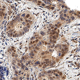
Detection of A20/TNFAIP3 in Human Colon. A20/TNFAIP3 was detected in immersion fixed paraffin-embedded sections of Human Colon using Mouse Anti-Human A20/TNFAIP3 Monoclonal Antibody (Catalog # MAB7598) at 5 µg/mL for 1 hour at room temperature followed by incubation with the Anti-Mouse IgG VisUCyte™ HRP Polymer Antibody (Catalog # VC001). Before incubation with the primary antibody, tissue was subjected to heat-induced epitope retrieval using VisUCyte Antigen Retrieval Reagent-Basic (Catalog # VCTS021). Tissue was stained using DAB (brown) and counterstained with hematoxylin (blue). Specific staining was localized to cytoplasm in epithelial cells in mucosal glands. View our protocol for IHC Staining with VisUCyte HRP Polymer Detection Reagents.
Reconstitution Calculator
Preparation and Storage
- 12 months from date of receipt, -20 to -70 °C as supplied.
- 1 month, 2 to 8 °C under sterile conditions after reconstitution.
- 6 months, -20 to -70 °C under sterile conditions after reconstitution.
Background: A20/TNFAIP3
A20 (TNF alpha -induced protein 3) is a cytoplasmic zinc finger protein that inhibits NF kappa B activity and tumor necrosis factor-mediated programmed cell death. The protein interacts with NAF1 and inhibits TNF-induced NF kappa B-dependent gene expression by interfering with RIP- or TRAF2-mediated transactivation signaling. A20 contains an N-terminal domain which has deubiquitinating enzyme activity and removes ubiquitin chains from receptor-interacting protein (RIP), thus mediating distinct regulatory effects in the down-regulation of NF kappa B signaling.
Product Datasheets
FAQs
No product specific FAQs exist for this product, however you may
View all Antibody FAQsReviews for Human A20/TNFAIP3 Antibody
There are currently no reviews for this product. Be the first to review Human A20/TNFAIP3 Antibody and earn rewards!
Have you used Human A20/TNFAIP3 Antibody?
Submit a review and receive an Amazon gift card.
$25/€18/£15/$25CAN/¥75 Yuan/¥2500 Yen for a review with an image
$10/€7/£6/$10 CAD/¥70 Yuan/¥1110 Yen for a review without an image

