Human A33 PE-conjugated Antibody Summary
Ile22-Val235
Accession # Q99795
Applications
Please Note: Optimal dilutions should be determined by each laboratory for each application. General Protocols are available in the Technical Information section on our website.
Scientific Data
 View Larger
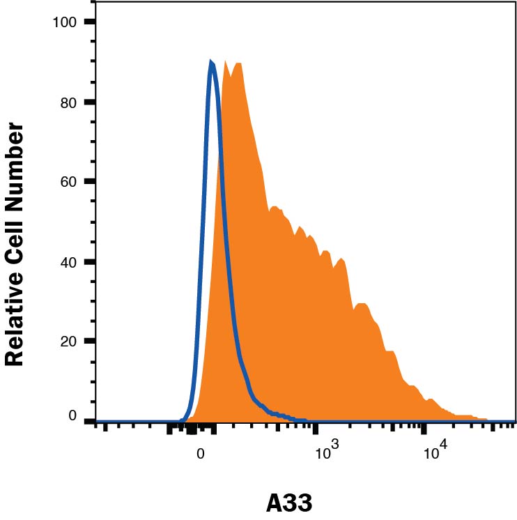
Detection of A33 in HT‑29 Human Cell Line by Flow Cytometry. HT-29 human colon adenocarcinoma cell line was stained with Rat Anti-Human A33 PE-conjugated Monoclonal Antibody (Catalog # FAB3080P, filled histogram) or isotype control antibody (Catalog # IC006P, open histogram). View our protocol for Staining Membrane-associated Proteins.
 View Larger
Detection of A33 in Colo-205 Human Cell Line by Flow Cytometry. Colo-205 human colon adenocarcinoma cell line was stained with Rat Anti-Human A33 PE-conjugated Monoclonal Antibody (Catalog # FAB3080P, filled histogram) or isotype control antibody (IC006P, open histogram). Staining was performed using our Staining Membrane-associated Proteins protocol.
Reconstitution Calculator
Preparation and Storage
Background: A33
Human A33, also known as GPA33, is a 43 kDa type I transmembrane glycoprotein that belongs to the CTX (cortical thymocyte marker in Xenopus) family of cell adhesion molecules within the immunoglobulin superfamily. Other family members include CXADR, ESAM, BT-IgSF, CD2 and JAM-A-C. The extracellular domain (ECD) of human A33 is 214 amino acids (aa) in length and contains one V-type and one C2-type Ig-like domain. This ECD is 80%, 74% and 71% aa identical to canine, bovine and mouse A33 ECD, respectively. A33 is likely to be involved in cell-cell adhesion between epithelial cells.
Product Datasheets
FAQs
No product specific FAQs exist for this product, however you may
View all Antibody FAQsReviews for Human A33 PE-conjugated Antibody
There are currently no reviews for this product. Be the first to review Human A33 PE-conjugated Antibody and earn rewards!
Have you used Human A33 PE-conjugated Antibody?
Submit a review and receive an Amazon gift card.
$25/€18/£15/$25CAN/¥75 Yuan/¥2500 Yen for a review with an image
$10/€7/£6/$10 CAD/¥70 Yuan/¥1110 Yen for a review without an image

