Human Activin RIA/ALK-2 Antibody Summary
Asp23-Glu123
Accession # Q04771
Applications
Please Note: Optimal dilutions should be determined by each laboratory for each application. General Protocols are available in the Technical Information section on our website.
Scientific Data
 View Larger
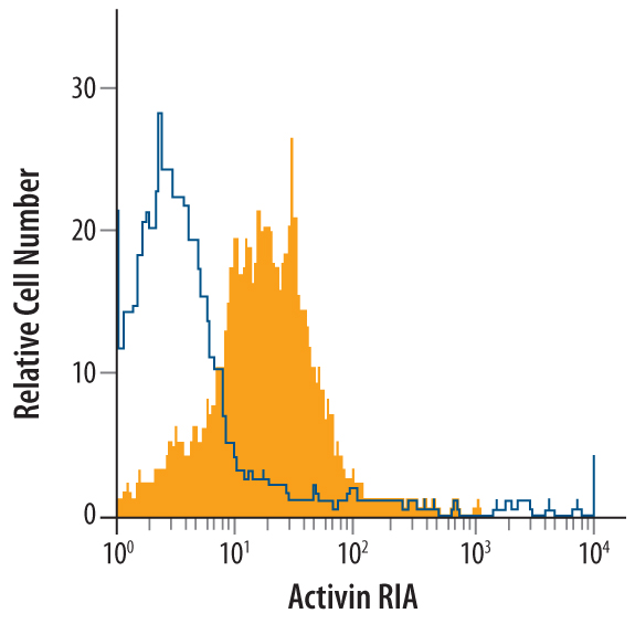
Detection of Activin RIA/ALK‑2 in PC‑3 Human Cell Line by Flow Cytometry. PC-3 human prostate cancer cell line was stained with Goat Anti-Human Activin RIA/ALK-2 Antigen Affinity-purified Polyclonal Antibody (Catalog # AF637, filled histogram) or control antibody (Catalog # AB-108-C, open histogram), followed by Phycoerythrin-conjugated Anti-Goat IgG Secondary Antibody (Catalog # F0107).
 View Larger
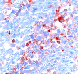
Activin RIA/ALK‑2 in Human Astrocytoma. Activin RIA/ALK-2 was detected in immersion fixed paraffin-embedded sections of human astrocytoma using 15 µg/mL Goat Anti-Human Activin RIA/ALK-2 Antigen Affinity-purified Polyclonal Antibody (Catalog # AF637) overnight at 4 °C. Tissue was stained using the Anti-Goat HRP-DAB Cell & Tissue Staining Kit (brown; Catalog # CTS008) and counterstained with hematoxylin (blue). View our protocol for Chromogenic IHC Staining of Paraffin-embedded Tissue Sections.
 View Larger
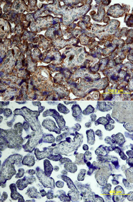
Activin RIA/ALK‑2 in Human Placenta. Activin RIA/ALK-2 was detected in immersion fixed paraffin-embedded sections of human placenta using Goat Anti-Human Activin RIA/ALK-2 Antigen Affinity-purified Polyclonal Antibody (Catalog # AF637) at 15 µg/mL overnight at 4 °C. Tissue was stained using the Anti-Goat HRP-DAB Cell & Tissue Staining Kit (brown; Catalog # CTS008) and counterstained with hematoxylin (blue). Lower panel shows a lack of labeling if primary antibodies are omitted and tissue is stained only with secondary antibody followed by incubation with detection reagents. View our protocol for Chromogenic IHC Staining of Paraffin-embedded Tissue Sections.
Reconstitution Calculator
Preparation and Storage
- 12 months from date of receipt, -20 to -70 °C as supplied.
- 1 month, 2 to 8 °C under sterile conditions after reconstitution.
- 6 months, -20 to -70 °C under sterile conditions after reconstitution.
Background: Activin RIA/ALK-2
Activin isoforms and other members of the TGF-beta superfamily exert their biological effects by binding to heteromeric complexes of a type I and a type II serine‑threonine kinase receptor, both of which are essential for signal transduction. To date, seven type I and five type II receptors, including the two type I and the two type II activin receptors, designated ActR-I(A), ActR-IB, ActR-II(A), and ActR-IIB, have been cloned from mammals. Through alternative mRNA splicing, multiple ActR-IIB isoforms can also be generated, adding to the complexity of the activin receptor system. Different activin isoforms bind with different high-affinities to the various type II isoforms. Type I activin receptors do not bind directly to activin but will associate with the type II receptor-activin complex and initiate signal transduction. Besides the activin isoforms, ActR-II will also bind inhibin, BMP-2 and BMP-7 with lower affinities. ActR-I can also bind and form signaling complexes with the BMP-2/7-bound BMPR-II. Activin type I receptors are highly conserved. Human, mouse and bovine type IA activin receptors share greater than 98% amino acid sequence homology. Recombinant soluble activin type I receptor does not bind activin.
- Attisano, L. et al. (1996) Mol. and Cell. Biol. 16:1066.
- Woodruff, T.K. (1998) Biochem. Pharmacology 55:953.
Product Datasheets
Citations for Human Activin RIA/ALK-2 Antibody
R&D Systems personnel manually curate a database that contains references using R&D Systems products. The data collected includes not only links to publications in PubMed, but also provides information about sample types, species, and experimental conditions.
4
Citations: Showing 1 - 4
Filter your results:
Filter by:
-
Chronic myeloid leukaemia cells require the bone morphogenic protein pathway for cell cycle progression and self-renewal
Authors: P Toofan, C Busch, H Morrison, S O'Brien, H Jørgensen, M Copland, H Wheadon
Cell Death Dis, 2018-09-11;9(9):927.
Species: Human
Sample Types: Whole Cells
Applications: Flow Cytometry -
Activation of Activin receptor-like kinases curbs mucosal inflammation and proliferation in chronic rhinosinusitis with nasal polyps
Authors: L Tengroth, J Arebro, O Larsson, C Bachert, SK Georén, LO Cardell
Sci Rep, 2018-01-24;8(1):1561.
Species: Human
Sample Types: Whole Cells
Applications: Flow Cytometry -
Establishment of a novel model of chondrogenesis using murine embryonic stem cells carrying fibrodysplasia ossificans progressiva-associated mutant ALK2.
Authors: Fujimoto M, Ohte S, Shin M, Yoneyama K, Osawa K, Miyamoto A, Tsukamoto S, Mizuta T, Kokabu S, Machiya A, Okuda A, Suda N, Katagiri T
Biochem Biophys Res Commun, 2014-11-15;455(3):347-52.
Species: Human
Sample Types: Whole Cells
Applications: Flow Cytometry -
EphB4 provides survival advantage to squamous cell carcinoma of the head and neck.
Authors: Masood R, Kumar SR, Sinha UK, Crowe DL, Krasnoperov V, Reddy RK, Zozulya S, Singh J, Xia G, Broek D, Schonthal AH, Gill PS
Int. J. Cancer, 2006-09-15;119(6):1236-48.
Species: Human
Sample Types: Cell Lysates
Applications: Western Blot
FAQs
No product specific FAQs exist for this product, however you may
View all Antibody FAQsReviews for Human Activin RIA/ALK-2 Antibody
There are currently no reviews for this product. Be the first to review Human Activin RIA/ALK-2 Antibody and earn rewards!
Have you used Human Activin RIA/ALK-2 Antibody?
Submit a review and receive an Amazon gift card.
$25/€18/£15/$25CAN/¥75 Yuan/¥2500 Yen for a review with an image
$10/€7/£6/$10 CAD/¥70 Yuan/¥1110 Yen for a review without an image

