Human Bad Antibody Summary
Met1-Gln168
Accession # Q92934
Applications
Please Note: Optimal dilutions should be determined by each laboratory for each application. General Protocols are available in the Technical Information section on our website.
Scientific Data
 View Larger
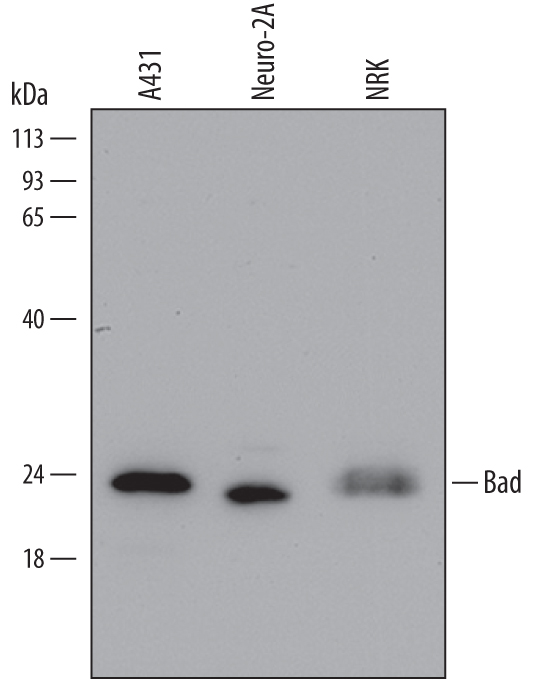
Detection of Human, Mouse, and Rat Bad by Western Blot. Western blot shows lysates of A431 human epithelial carcinoma cell line, Neuro-2A mouse neuroblastoma cell line, and NRK rat normal kidney cell line. PVDF Membrane was probed with 0.5 µg/mL of Mouse Anti-Human/Mouse/Rat Bad Monoclonal Antibody (Catalog # MAB6405) followed by HRP-conjugated Anti-Mouse IgG Secondary Antibody (Catalog # HAF007). A specific band was detected for Bad at approximately 23 kDa (as indicated). This experiment was conducted under reducing conditions and using Immunoblot Buffer Group 2.
 View Larger
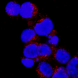
Bad in Jurkat Human Cell Line. Bad was detected in immersion fixed Jurkat human acute T cell leukemia cell line using Mouse Anti-Human Bad Monoclonal Antibody (Catalog # MAB6405) at 25 µg/mL for 3 hours at room temperature. Cells were stained using the NorthernLights™ 557-conjugated Anti-Mouse IgG Secondary Antibody (red; Catalog # NL007) and counterstained with DAPI (blue). Specific staining was localized to cytoplasm. View our protocol for Fluorescent ICC Staining of Non-adherent Cells.
 View Larger
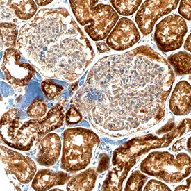
Bad in Human Kidney. Bad was detected in immersion fixed paraffin-embedded sections of human kidney using Mouse Anti-Human/Mouse/Rat Bad Monoclonal Antibody (Catalog # MAB6405) at 15 µg/mL overnight at 4 °C. Before incubation with the primary antibody, tissue was subjected to heat-induced epitope retrieval using Antigen Retrieval Reagent-Basic (Catalog # CTS013). Tissue was stained using the Anti-Mouse HRP-DAB Cell & Tissue Staining Kit (brown; Catalog # CTS002) and counterstained with hematoxylin (blue). Specific staining was localized to cytoplasm of epithelial cells in convoluted tubules. View our protocol for Chromogenic IHC Staining of Paraffin-embedded Tissue Sections.
 View Larger
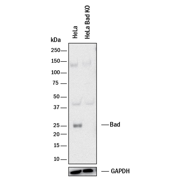
Western Blot Shows Human Bad Specificity by Using Knockout Cell Line. Western blot shows lysates of HeLa human cervical epithelial carcinoma parental cell line and Bad knockout HeLa cell line (KO). PVDF membrane was probed with 0.5 µg/mL of Mouse Anti-Human Bad Monoclonal Antibody (Catalog # MAB6405) followed by HRP-conjugated Anti-Mouse IgG Secondary Antibody (Catalog # HAF018). A specific band was detected for Bad at approximately 25 kDa (as indicated) in the parental HeLa cell line, but is not detectable in knockout HeLa cell line. GAPDH (Catalog # MAB5718) is shown as a loading control. This experiment was conducted under reducing conditions and using Immunoblot Buffer Group 1.
Reconstitution Calculator
Preparation and Storage
- 12 months from date of receipt, -20 to -70 °C as supplied.
- 1 month, 2 to 8 °C under sterile conditions after reconstitution.
- 6 months, -20 to -70 °C under sterile conditions after reconstitution.
Background: Bad
Bcl-2 antagonist of cell death (Bad) is a 23 kDa cytoplasmic protein in the Bcl-2 family. It functions as a pro-apoptotic molecule by dimerizing with and inhibiting the anti-apoptotic proteins Bcl-2 and Bcl-xL. Prosurvival signals trigger the phosphorylation of Bad on Ser115, disrupting its interaction with Bcl-2 and Bcl-xL and resulting in protection from apoptosis. Human Bad shares 75% aa sequence identity with mouse and rat Bad.
Product Datasheets
Citation for Human Bad Antibody
R&D Systems personnel manually curate a database that contains references using R&D Systems products. The data collected includes not only links to publications in PubMed, but also provides information about sample types, species, and experimental conditions.
1 Citation: Showing 1 - 1
-
FK506 Attenuates the MRP1-Mediated Chemoresistant Phenotype in Glioblastoma Stem-Like Cells
Authors: Á Torres, V Arriagada, JI Erices, MLÁ Toro, JD Rocha, I Niechi, C Carrasco, C Oyarzún, C Quezada
Int J Mol Sci, 2018-09-11;19(9):.
Species: Human
Sample Types: Cell Lysates
Applications: Western Blot
FAQs
No product specific FAQs exist for this product, however you may
View all Antibody FAQsReviews for Human Bad Antibody
Average Rating: 5 (Based on 2 Reviews)
Have you used Human Bad Antibody?
Submit a review and receive an Amazon gift card.
$25/€18/£15/$25CAN/¥75 Yuan/¥2500 Yen for a review with an image
$10/€7/£6/$10 CAD/¥70 Yuan/¥1110 Yen for a review without an image
Filter by:
Antibody was printed on custom arrays and incubated with fluorescently labeled human EDTA plasma


