Human BMI-1 Antibody Summary
Asp96-Gly326
Accession # P35226
Applications
Please Note: Optimal dilutions should be determined by each laboratory for each application. General Protocols are available in the Technical Information section on our website.
Scientific Data
 View Larger
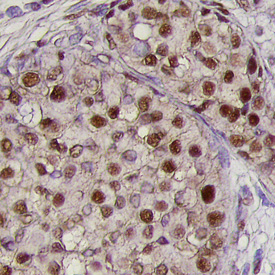
BMI‑1 in Human Breast Cancer Tissue. BMI‑1 was detected in immersion fixed paraffin-embedded sections of human breast cancer tissue using Human BMI‑1 Monoclonal Antibody (Catalog # MAB33342) at 25 µg/mL overnight at 4 °C. Tissue was stained using the Anti-Mouse HRP-DAB Cell & Tissue Staining Kit (brown; Catalog # CTS002) and counterstained with hematoxylin (blue). Specific labeling was localized to the nuclei of epithelial cells. View our protocol for Chromogenic IHC Staining of Paraffin-embedded Tissue Sections.
 View Larger
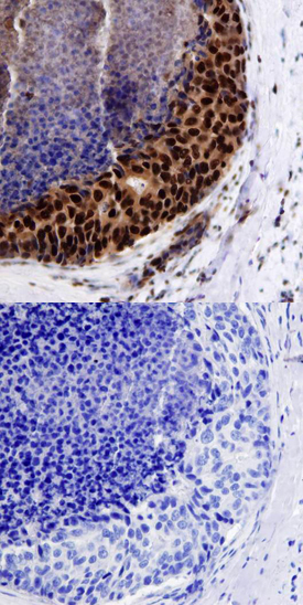
BMI‑1 in Human Breast. BMI-1 was detected in immersion fixed paraffin-embedded sections of normal human breast using Human BMI-1 Monoclonal Antibody (Catalog # MAB33342) at 15 µg/mL overnight at 4 °C. Tissue was stained using the Anti-Mouse HRP-DAB Cell & Tissue Staining Kit (brown; Catalog # CTS002) and counterstained with hematoxylin (blue). Lower panel shows a lack of labeling if primary antibodies are omitted and tissue is stained only with secondary antibody followed by incubation with detection reagents. View our protocol for Chromogenic IHC Staining of Paraffin-embedded Tissue Sections.
Reconstitution Calculator
Preparation and Storage
- 12 months from date of receipt, -20 to -70 °C as supplied.
- 1 month, 2 to 8 °C under sterile conditions after reconstitution.
- 6 months, -20 to -70 °C under sterile conditions after reconstitution.
Background: BMI-1
BMI-1 (B cell-specific Moloney-MLV integration site #1) is a 45 kDa protooncogene that is a class II member of the Polycomb group of genes. It participates in the formation of a large multimeric complex termed PRC1 that inhibits target gene transcription. Loss of BMI-1 function precludes stem cells from self-replicating. Human BMI-1 contains an N-terminal RING-finger domain (aa 17-56), an NLS (aa 81-95) and a C-terminal Pro/Ser-rich region (aa 251-326). Human BMI-1 is 99%, 97%, 99% and 99% aa identical to bovine, mouse, feline and canine BMI-1, respectively.
Product Datasheets
Citations for Human BMI-1 Antibody
R&D Systems personnel manually curate a database that contains references using R&D Systems products. The data collected includes not only links to publications in PubMed, but also provides information about sample types, species, and experimental conditions.
3
Citations: Showing 1 - 3
Filter your results:
Filter by:
-
Lymphocyte to monocyte ratio predicts survival and is epigenetically linked to miR-222-3p and miR-26b-5p in diffuse large B cell lymphoma
Authors: AM Metwally, AAHM Kasem, MI Youssif, SM Hassan, AHA Abdel Waha, LA Refaat
Scientific Reports, 2023-03-25;13(1):4899.
Species: Human
Sample Types: Whole Tissue
Applications: IHC -
FUN14 domain-containing 1 promotes breast cancer proliferation and migration by activating calcium-NFATC1-BMI1 axis
Authors: L Wu, D Zhang, L Zhou, Y Pei, Y Zhuang, W Cui, J Chen
EBioMedicine, 2019-02-23;0(0):.
Species: Human
Sample Types: Cell Lysates, Whole Tissue
Applications: IHC-P, Western Blot -
p53 regulates epithelial-mesenchymal transition and stem cell properties through modulating miRNAs.
Authors: Chang CJ, Chao CH, Xia W, Yang JY, Xiong Y, Li CW, Yu WH, Rehman SK, Hsu JL, Lee HH, Liu M, Chen CT, Yu D, Hung MC
Nat. Cell Biol., 2011-02-20;13(3):317-23.
Species: Human
Sample Types: Whole Tissue
Applications: IHC
FAQs
No product specific FAQs exist for this product, however you may
View all Antibody FAQsReviews for Human BMI-1 Antibody
There are currently no reviews for this product. Be the first to review Human BMI-1 Antibody and earn rewards!
Have you used Human BMI-1 Antibody?
Submit a review and receive an Amazon gift card.
$25/€18/£15/$25CAN/¥75 Yuan/¥2500 Yen for a review with an image
$10/€7/£6/$10 CAD/¥70 Yuan/¥1110 Yen for a review without an image


