Human Carboxypeptidase E/CPE Antibody Summary
Arg42-Ser453
Accession # P16870
Applications
Please Note: Optimal dilutions should be determined by each laboratory for each application. General Protocols are available in the Technical Information section on our website.
Scientific Data
 View Larger
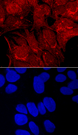
Carboxypeptidase E/CPE in HepG2 Human Cell Line. Carboxypeptidase E/CPE was detected in immersion fixed HepG2 human hepatocellular carcinoma cell line using Goat Anti-Human Carboxypeptidase E/CPE Antigen Affinity-purified Polyclonal Antibody (Catalog # AF3587) at 10 µg/mL for 3 hours at room temperature. Cells were stained using the NorthernLights™ 557-conjugated Anti-Goat IgG Secondary Antibody (red, upper panel; Catalog # NL001) and counterstained with DAPI (blue, lower panel). Specific staining was localized to cell surfaces and cytoplasm. View our protocol for Fluorescent ICC Staining of Cells on Coverslips.
 View Larger
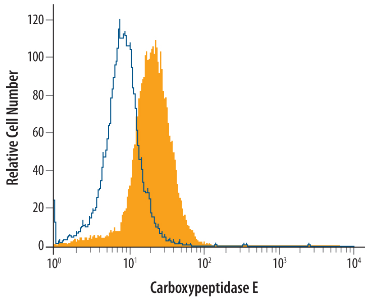
Detection of Carboxypeptidase E/CPE in A172 Human Cell Line by Flow Cytometry. A172 human glioblastoma cell line was stained with Goat Anti-Human Carboxypeptidase E/CPE Antigen Affinity-purified Polyclonal Antibody (Catalog # AF3587, filled histogram) or control antibody (Catalog # AB-108-C, open histogram), followed by Phycoerythrin-conjugated Anti-Goat IgG Secondary Antibody (Catalog # F0107). To facilitate intracellular staining, cells were fixed with paraformaldehyde and permeabilized with methanol and saponin.
 View Larger
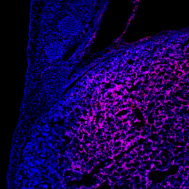
Carboxypeptidase E/CPE in Mouse Embryo. Carboxypeptidase E/CPE was detected in immersion fixed frozen sections of mouse embryo (9.5 d.p.c.) using Goat Anti-Human Carboxypeptidase E/CPE Antigen Affinity-purified Polyclonal Antibody (Catalog # AF3587) at 10 µg/mL overnight at 4 °C. Tissue was stained using the NorthernLights™ 557-conjugated Anti-Goat IgG Secondary Antibody (red; Catalog # NL001) and counterstained with DAPI (blue). Specific staining was localized to the developing liver. View our protocol for Fluorescent IHC Staining of Frozen Tissue Sections.
Reconstitution Calculator
Preparation and Storage
- 12 months from date of receipt, -20 to -70 °C as supplied.
- 1 month, 2 to 8 °C under sterile conditions after reconstitution.
- 6 months, -20 to -70 °C under sterile conditions after reconstitution.
Background: Carboxypeptidase E/CPE
Encoded by the CPE gene and also known as Carboxypeptidase H, CPE is a single chain peptidase with an optimal pH range between 5.0‑6.0. It is a zinc metallocarboxypeptidase that removes basic amino acids from the C-terminus of peptides (1). Like other metallocarboxypeptidases, its activity is stimulated by millimolar concentrations of Co2+. Its activity is regulated by pH-induced aggregation above pH 6.0. Its major function seems to process numerous peptide hormones and neurotransmitters. In addition to its proteolytic function, it also plays a role as a sorting receptor (2), which may be attributed to the sorting of this protein into the secretory pathway. The C-terminal domain of CPE causes the peripheral association of CPE with membranes below neutral pH, resulting in the association of this protein into membranes (3). CPE knockout mice live but become obese due to impaired glucose clearance and insulin resistance (4).
- Fricker, L.D. (2004) in Handbook of Proteolytic Enzymes, Barrett, A.J. et al. eds. pp. 840.
- Cool, D.R. et al. (1997) Cell 88:73.
- Zhang, C-F. et al. (2003) Biochem. J. 369:453.
- Cawley, N.X. et al. (2004) Endocrinology 145:5807.
Product Datasheets
Citation for Human Carboxypeptidase E/CPE Antibody
R&D Systems personnel manually curate a database that contains references using R&D Systems products. The data collected includes not only links to publications in PubMed, but also provides information about sample types, species, and experimental conditions.
1 Citation: Showing 1 - 1
-
Loss of MAGEL2 in Prader-Willi syndrome leads to decreased secretory granule and neuropeptide production
Authors: H Chen, AK Victor, J Klein, KF Tacer, DJ Tai, C de Esch, A Nuttle, J Temirov, LC Burnett, M Rosenbaum, Y Zhang, L Ding, JJ Moresco, JK Diedrich, JR Yates, HS Tillman, RL Leibel, ME Talkowski, DD Billadeau, LT Reiter, PR Potts
JCI Insight, 2020-09-03;5(17):.
Species: Mouse
Sample Types: Cell Lysates, Whole Cells
Applications: ICC, Western Blot
FAQs
No product specific FAQs exist for this product, however you may
View all Antibody FAQsReviews for Human Carboxypeptidase E/CPE Antibody
There are currently no reviews for this product. Be the first to review Human Carboxypeptidase E/CPE Antibody and earn rewards!
Have you used Human Carboxypeptidase E/CPE Antibody?
Submit a review and receive an Amazon gift card.
$25/€18/£15/$25CAN/¥75 Yuan/¥2500 Yen for a review with an image
$10/€7/£6/$10 CAD/¥70 Yuan/¥1110 Yen for a review without an image

