Human CCL4/MIP-1 beta Fluorescein-conjugated Antibody Summary
Ala24-Asn92
Accession # P13236
Applications
Please Note: Optimal dilutions should be determined by each laboratory for each application. General Protocols are available in the Technical Information section on our website.
Scientific Data
 View Larger
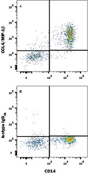
Detection of CCL4/MIP‑1 beta in Human Blood Monocytes by Flow Cytometry. Human peripheral blood monocytes treated with LPS and Monensin for 5 hours were stained with Mouse Anti-Human CD14 APC-conjugated Monoclonal Antibody (Catalog # FAB3832A) and either (A) Mouse Anti-Human CCL4/MIP-1 beta Fluorescein-conjugated Monoclonal Antibody (Catalog # IC271F) or (B) Mouse IgG2BFluorescein Isotype Control (Catalog # IC0041F). To facilitate intracellular staining, cells were fixed with Flow Cytometry Fixation Buffer (Catalog # FC004) and permeabilized with Flow Cytometry Permeabilization/Wash Buffer I (Catalog # FC005). View our protocol for Staining Intracellular Molecules.
Reconstitution Calculator
Preparation and Storage
Background: CCL4/MIP-1 beta
CCL4, also known as Macrophage Inflammatory Protein 1 beta (MIP-1 beta ) is a member of the CC or beta chemokine subfamily. CCL4 is expressed primarily by T cells, B cells, and monocytes after antigen or mitogen stimulation. The functional receptor for CCL4 has been identified as CCR5. Mature human CCL4 shares 77% and 80% aa sequence identity with mouse and rat CCL4, respectively.
Product Datasheets
Citations for Human CCL4/MIP-1 beta Fluorescein-conjugated Antibody
R&D Systems personnel manually curate a database that contains references using R&D Systems products. The data collected includes not only links to publications in PubMed, but also provides information about sample types, species, and experimental conditions.
12
Citations: Showing 1 - 10
Filter your results:
Filter by:
-
Preservation of Lymphopoietic Potential and Virus Suppressive Capacity by CD8+ T Cells in HIV-2-Infected Controllers
J Immunol, 2016-08-26;0(0):.
Species: Human
Sample Types: Whole Cells
Applications: Flow Cytometry -
Human CD8+ T-cells recognizing peptides from Mycobacterium tuberculosis (Mtb) presented by HLA-E have an unorthodox Th2-like, multifunctional, Mtb inhibitory phenotype and represent a novel human T-cell subset.
Authors: van Meijgaarden K, Haks M, Caccamo N, Dieli F, Ottenhoff T, Joosten S
PLoS Pathog, 2015-03-24;11(3):e1004671.
Species: Human
Sample Types: Whole Cells
Applications: Flow Cytometry -
CD8+ regulatory T cells, and not CD4+ T cells, dominate suppressive phenotype and function after in vitro live Mycobacterium bovis-BCG activation of human cells.
Authors: Boer M, van Meijgaarden K, Joosten S, Ottenhoff T
PLoS ONE, 2014-04-08;9(4):e94192.
Species: Human
Sample Types: Whole Cells
Applications: Flow Cytometry -
CD39 is involved in mediating suppression by Mycobacterium bovis BCG-activated human CD8(+) CD39(+) regulatory T cells.
Authors: Boer M, van Meijgaarden K, Bastid J, Ottenhoff T, Joosten S
Eur J Immunol, 2013-05-29;43(7):1925-32.
Species: Human
Sample Types: Whole Cells
Applications: Flow Cytometry -
A molecular basis for the control of preimmune escape variants by HIV-specific CD8+ T cells.
Authors: Ladell K, Hashimoto M, Iglesias M, Wilmann P, McLaren J, Gras S, Chikata T, Kuse N, Fastenackels S, Gostick E, Bridgeman J, Venturi V, Arkoub Z, Agut H, van Bockel D, Almeida J, Douek D, Meyer L, Venet A, Takiguchi M, Rossjohn J, Price D, Appay V
Immunity, 2013-03-21;38(3):425-36.
Species: Human
Sample Types: Whole Cells
Applications: Flow Cytometry -
Rapid antigen processing and presentation of a protective and immunodominant HLA-B*27-restricted hepatitis C virus-specific CD8+ T-cell epitope.
Authors: Schmidt J, Iversen A, Tenzer S, Gostick E, Price D, Lohmann V, Distler U, Bowness P, Schild H, Blum H, Klenerman P, Neumann-Haefelin C, Thimme R
PLoS Pathog, 2012-11-29;8(11):e1003042.
Species: Human
Sample Types: Whole Cells
Applications: Flow Cytometry -
Early Induction of Polyfunctional Simian Immunodeficiency Virus (SIV)-Specific T Lymphocytes and Rapid Disappearance of SIV from Lymph Nodes of Sooty Mangabeys during Primary Infection.
Authors: Meythaler M, Wang Z, Martinot A, Pryputniewicz S, Kasheta M, McClure HM, O'Neil SP, Kaur A
J. Immunol., 2011-03-25;186(9):5151-61.
Species: Primate - Cercocebus torquatus (Sooty Mangabey)
Sample Types: Whole Cells
Applications: Flow Cytometry -
Interleukin-27 induces a STAT1/3- and NF-kappaB-dependent proinflammatory cytokine profile in human monocytes.
Authors: Guzzo C, Che Mat NF, Gee K
J. Biol. Chem., 2010-06-02;285(32):24404-11.
Species: Human
Sample Types: Whole Cells
Applications: Flow Cytometry -
CD16- natural killer cells: enrichment in mucosal and secondary lymphoid tissues and altered function during chronic SIV infection.
Authors: Reeves RK, Gillis J, Wong FE
Blood, 2010-03-25;115(22):4439-46.
Species: Primate - Macaca mulatta (Rhesus Macaque)
Sample Types: Whole Cells
Applications: Flow Cytometry -
Comprehensive assessment of chemokine expression profiles by flow cytometry.
Authors: Eberlein J, Nguyen TT, Victorino F, Golden-Mason L, Rosen HR, Homann D
J. Clin. Invest., 2010-02-08;120(3):907-23.
Species: Human
Sample Types: Whole Cells
Applications: Flow Cytometry -
Cognate CD4+ T-cell-dendritic cell interactions induce migration of immature dendritic cells through dissolution of their podosomes.
Authors: Nobile C, Lind M, Miro F, Chemin K, Tourret M, Occhipinti G, Dogniaux S, Amigorena S, Hivroz C
Blood, 2008-01-18;111(7):3579-90.
Species: Human
Sample Types: Whole Cells
Applications: Flow Cytometry -
Chemokines and their receptors in whiplash injury: elevated RANTES and CCR-5.
Authors: Kivioja J, Rinaldi L, Ozenci V, Kouwenhoven M, Kostulas N, Lindgren U, Link H
J. Clin. Immunol., 2001-07-01;21(4):272-7.
Species: Human
Sample Types: Whole Cells
Applications: Flow Cytometry
FAQs
No product specific FAQs exist for this product, however you may
View all Antibody FAQsReviews for Human CCL4/MIP-1 beta Fluorescein-conjugated Antibody
There are currently no reviews for this product. Be the first to review Human CCL4/MIP-1 beta Fluorescein-conjugated Antibody and earn rewards!
Have you used Human CCL4/MIP-1 beta Fluorescein-conjugated Antibody?
Submit a review and receive an Amazon gift card.
$25/€18/£15/$25CAN/¥75 Yuan/¥2500 Yen for a review with an image
$10/€7/£6/$10 CAD/¥70 Yuan/¥1110 Yen for a review without an image


