Human CCR9 Antibody
Human CCR9 Antibody Summary
Met1-Leu369
Accession # P51686
Applications
Please Note: Optimal dilutions should be determined by each laboratory for each application. General Protocols are available in the Technical Information section on our website.
Scientific Data
 View Larger
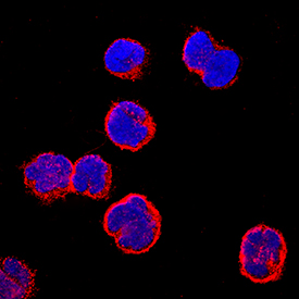
CCR9 in MOLT‑4 Human Cell Line. CCR9 was detected in immersion fixed MOLT-4 human acute lymphoblastic leukemia cell line using Mouse Anti-Human CCR9 Monoclonal Antibody (Catalog # MAB179) at 25 µg/mL for 3 hours at room temperature. Cells were stained using the NorthernLights™ 557-conjugated Anti-Mouse IgG Secondary Antibody (red; Catalog # NL007) and counterstained with DAPI (blue). Specific staining was localized to plasma membranes. View our protocol for Fluorescent ICC Staining of Non-adherent Cells.
Reconstitution Calculator
Preparation and Storage
- 12 months from date of receipt, -20 to -70 °C as supplied.
- 1 month, 2 to 8 °C under sterile conditions after reconstitution.
- 6 months, -20 to -70 °C under sterile conditions after reconstitution.
Background: CCR9
CCR9 is a G protein-linked seven transmembrane domain cytokine receptor. CCR9 serves as a receptor for CCL25/TECK. It is expressed on mature and immature thymocytes and some peripheral T and B cells.
Product Datasheets
Citations for Human CCR9 Antibody
R&D Systems personnel manually curate a database that contains references using R&D Systems products. The data collected includes not only links to publications in PubMed, but also provides information about sample types, species, and experimental conditions.
8
Citations: Showing 1 - 8
Filter your results:
Filter by:
-
Novel peptide screened from a phage display library antagonizes the activity of CC chemokine receptor 9
Authors: Y Hu, A Ma, S Lin, Y Yang, G Hong
Oncol Lett, 2017-09-26;14(6):6471-6476.
Species: Human
Sample Types: Whole Cells
Applications: ICC -
Intracellular allosteric antagonism of the CCR9 receptor
Nature, 2016-12-07;540(7633):462-465.
Species: Human
Sample Types: Whole Cells
Applications: Flow Cytometry -
CCL25 mediates migration, invasion and matrix metalloproteinase expression by breast cancer cells in a CCR9-dependent fashion.
Authors: Johnson-Holiday C, Singh R, Johnson E
Int. J. Oncol., 2011-02-23;38(5):1279-85.
Species: Human
Sample Types: Whole Cells
Applications: Neutralization -
Distribution, persistence, and efficacy of adoptively transferred central and effector memory-derived autologous Simian Immunodeficiency Virus-specific CD8(+) T cell clones in rhesus macaques during acute infection.
Authors: Minang JT, Trivett MT, Bolton DL, Trubey CM, Estes JD, Li Y, Smedley J, Pung R, Rosati M, Jalah R, Pavlakis GN, Felber BK, Piatak M, Roederer M, Lifson JD, Ott DE, Ohlen C
J. Immunol., 2009-11-30;184(1):315-26.
Species: Primate - Macaca mulatta (Rhesus Macaque)
Sample Types: Whole Cells
Applications: Flow Cytometry -
Trafficking, persistence, and activation state of adoptively transferred allogeneic and autologous Simian Immunodeficiency Virus-specific CD8(+) T cell clones during acute and chronic infection of rhesus macaques.
Authors: Bolton DL, Minang JT, Trivett MT, Song K, Tuscher JJ, Li Y, Piatak M, O'Connor D, Lifson JD, Roederer M, Ohlen C
J. Immunol., 2009-11-30;184(1):303-14.
Species: Primate - Macaca mulatta (Rhesus Macaque)
Sample Types: Whole Cells
Applications: Flow Cytometry -
Down-modulation of CXCR3 surface expression and function in CD8+ T cells from cutaneous T cell lymphoma patients.
Authors: Winter D, Moser J, Kriehuber E, Wiesner C, Knobler R, Trautinger F, Bombosi P, Stingl G, Petzelbauer P, Rot A, Maurer D
J. Immunol., 2007-09-15;179(6):4272-82.
Species: Human
Sample Types: Whole Cells
Applications: Flow Cytometry -
The monoclonal antibody CHO-131 identifies a subset of cutaneous lymphocyte-associated antigen T cells enriched in P-selectin-binding cells.
Authors: Ni Z, Campbell JJ, Niehans G, Walcheck B
J. Immunol., 2006-10-01;177(7):4742-8.
Species: Human
Sample Types: Whole Cells
Applications: Flow Cytometry -
Possible link between unique chemokine and homing receptor expression at diagnosis and relapse location in a patient with childhood T-ALL.
Authors: Annels NE, Willemze AJ, van der Velden VH, Faaij CM, van Wering E, Sie-Go DM, Egeler RM, van Tol MJ, Révész T
Blood, 2003-12-04;103(7):2806-8.
Species: Human
Sample Types: Whole Cells
Applications: Flow Cytometry
FAQs
No product specific FAQs exist for this product, however you may
View all Antibody FAQsReviews for Human CCR9 Antibody
Average Rating: 5 (Based on 1 Review)
Have you used Human CCR9 Antibody?
Submit a review and receive an Amazon gift card.
$25/€18/£15/$25CAN/¥75 Yuan/¥2500 Yen for a review with an image
$10/€7/£6/$10 CAD/¥70 Yuan/¥1110 Yen for a review without an image
Filter by:
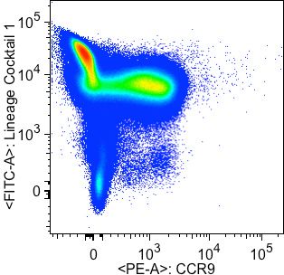
Gated on lymphocyte FSC-A and SSC-A populations. Singlets were further gated on by FSC-A/FSC-H doublet exclusion. Lineage Cocktail 1 (BD Biosciences) comprises CD3, CD14, CD16, CD19, CD20, and CD56 antibodies.
Specificity: Specific
Sensitivity: Sensitive
Buffer: 2% Human Serum, 0.5 mM EDTA in PBS
Dilution: 1/100


