Human CD30/TNFRSF8 Antibody Summary
Phe19-Lys379
Accession # P28908
Applications
under non-reducing conditions only
0.05 - 0.2 μg/mL.
Please Note: Optimal dilutions should be determined by each laboratory for each application. General Protocols are available in the Technical Information section on our website.
Scientific Data
 View Larger
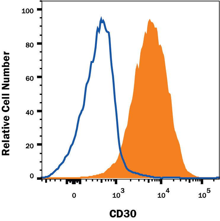
Detection of CD30/TNFRSF8 in Jurkat Human Cell Line by Flow Cytometry. Jurkat human acute T cell leukemia cell line was stained with Mouse Anti-Human CD30/TNFRSF8 Monoclonal Antibody (Catalog # MAB229, filled histogram) or isotype control antibody (Catalog # MAB0041, open histogram), followed by Phycoerythrin-conjugated Anti-Mouse IgG Secondary Antibody (Catalog # F0102B). View our protocol for Staining Membrane-associated Proteins.
 View Larger
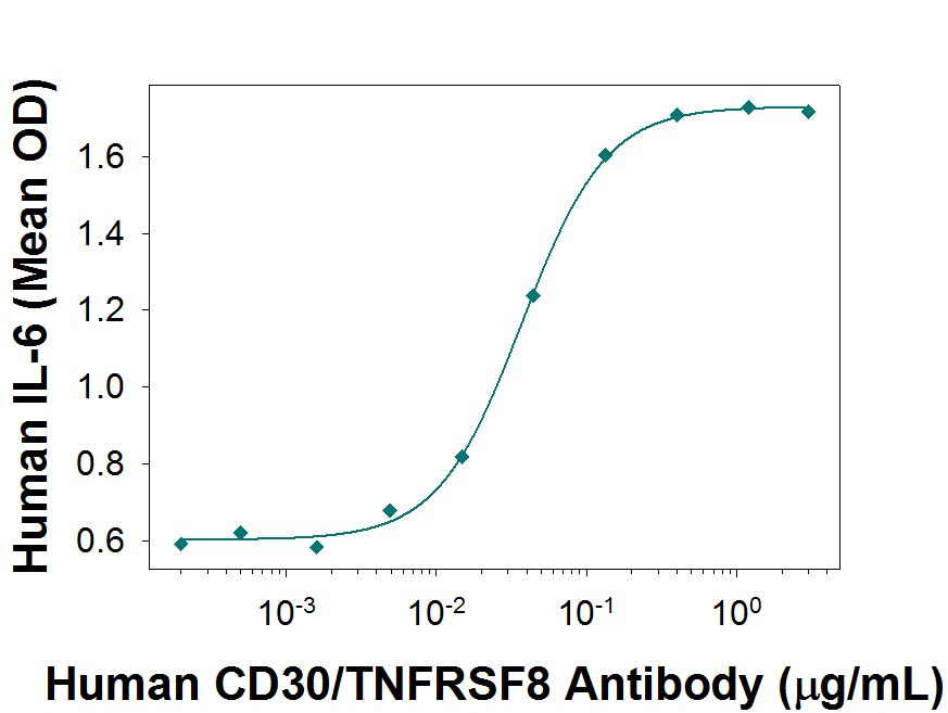
Human CD30/TNFRSF8 Antibody Enhances IL-6 Secretion in HDLM-2 Cells. Human CD30/TNFRSF8 Monoclonal Antibody enhances IL-6 secretion in the HDLM-2 human Hodgkin's lymphoma cell line, in a dose-dependent manner, as measured using the Quantikine Human IL-6 ELISA Kit (Catalog # D6050). The ED50 for this effect is typically 0.05- 0.2 µg/mL.
Reconstitution Calculator
Preparation and Storage
- 12 months from date of receipt, -20 to -70 °C as supplied.
- 1 month, 2 to 8 °C under sterile conditions after reconstitution.
- 6 months, -20 to -70 °C under sterile conditions after reconstitution.
Background: CD30/TNFRSF8
CD30, also known as Ki-1 antigen and TNFRSF8, is a 120 kDa type I transmembrane glycoprotein belonging to the TNF receptor superfamily (1, 2). Mature human CD30 consists of a 361 amino acid (aa) extracellular domain (ECD) with six cysteine-rich repeats, a 28 aa transmembrane segment, and a 188 aa cytoplasmic domain (3). In contrast, mouse and rat CD30 lack 90 aa of the ECD and contain only three cysteine-rich repeats. Within common regions of the ECD, human CD30 shares 53% and 49% aa sequence identity with mouse and rat CD30, respectively. Alternate splicing of human CD30 generates an isoform that includes only the C‑terminal 132 aa of the cytoplasmic domain. CD30 is normally expressed on antigen-stimulated Th cells and B cells (4 - 6). However, it is upregulated in Hodgkin’s disease (on Reed-Sternberg cells), other lymphomas, chronic inflammation, and autoimmunity (7). CD30 binds to CD30 Ligand/TNFSF8 which is expressed on activated Th cells, monocytes, granulocytes and medullary thymic epithelial cells (1, 5). CD30 signaling costimulates antigen-induced Th0 and Th2 proliferation and cytokine secretion but favors a Th2-biased immune response (8). In the absence of antigenic stimulation, it can still induce T cell expression of IL-13 (9). CD30 contributes to thymic negative selection by inducing the apoptotic cell death of CD4+CD8+ T cells (10, 11). In B cells, CD30 ligation promotes cellular proliferation and antibody production in addition to the expression of CXCR4, CCL3, and CCL5 (5, 12). An 85-90 kDa soluble form of CD30 is shed from the cell surface by TACE-mediated cleavage (13, 14). Soluble CD30 retains the ability to bind CD30 Ligand and functions as an inhibitor of normal CD30 signaling (15).
- Kennedy, M.K. et al. (2006) Immunology 118:143.
- Tarkowski, M. (2003) Curr. Opin. Hematol. 10:267.
- Durkop, H. et al. (1992) Cell 68:421.
- Hamann, D. et al. (1996) J. Immunol. 156:1387.
- Shanebeck, S.D. et al. (1995) Eur. J. Immunol. 25:2147.
- Gruss, H.-J. et al. (1994) Blood 83:2045.
- Oflzoglu E. et al. (2009) Adv. Exp. Med. Biol. 647:174.
- Del Prete, G. et al. (1995) J. Exp. Med. 182:1655.
- Harlin, H. et al. (2002) J. Immunol. 169:2451.
- Amakawa, R. et al. (1996) Cell 84:551.
- Chiarle, R. et al. (1999) J. Immunol. 163:194.
- Vinante, F. et al. (2002) Blood 99:52.
- Hansen, H.P. et al. (1995) Int. J. Cancer 63:750.
- Hansen, H.P. et al. (2000) J. Immunol. 165:6703.
- Hargreaves, P.G. and A. Al-Shamkhani (2002) Eur. J. Immunol. 32:163.
Product Datasheets
Citations for Human CD30/TNFRSF8 Antibody
R&D Systems personnel manually curate a database that contains references using R&D Systems products. The data collected includes not only links to publications in PubMed, but also provides information about sample types, species, and experimental conditions.
3
Citations: Showing 1 - 3
Filter your results:
Filter by:
-
Molecular Signatures of Dengue Virus-Specific IL-10/IFN-gamma Co-producing CD4�T Cells and Their Association with Dengue Disease
Authors: Y Tian, G Seumois, LM De-Oliveir, J Mateus, S Herrera-de, C Kim, D Hinz, NDS Goonawardh, AD de Silva, S Premawansa, G Premawansa, A Wijewickra, A Balmaseda, A Grifoni, P Vijayanand, E Harris, B Peters, A Sette, D Weiskopf
Cell Rep, 2019-12-24;29(13):4482-4495.e4.
Species: Human
Sample Types: Whole Cells
Applications: Flow Cytometry -
Cytoplasmic Pin1 expression is increased in human cutaneous melanoma and predicts poor prognosis
Authors: X Chen, X Liu, B Deng, M Martinka, Y Zhou, X Lan, Y Cheng
Sci Rep, 2018-11-15;8(1):16867.
Species: Human
Sample Types: Cell Lysates, Tissue Array
Applications: IHC-P, Western Blot -
Selective redox regulation of cytokine receptor signaling by extracellular thioredoxin-1.
Authors: Schwertassek U, Balmer Y, Gutscher M, Weingarten L, Preuss M, Engelhard J, Winkler M, Dick TP
EMBO J., 2007-06-07;26(13):3086-97.
Species: Human
Sample Types: Whole Cells
Applications: Flow Cytometry
FAQs
No product specific FAQs exist for this product, however you may
View all Antibody FAQsReviews for Human CD30/TNFRSF8 Antibody
There are currently no reviews for this product. Be the first to review Human CD30/TNFRSF8 Antibody and earn rewards!
Have you used Human CD30/TNFRSF8 Antibody?
Submit a review and receive an Amazon gift card.
$25/€18/£15/$25CAN/¥75 Yuan/¥2500 Yen for a review with an image
$10/€7/£6/$10 CAD/¥70 Yuan/¥1110 Yen for a review without an image


