Human CD36/SR-B3 Antibody Summary
Applications
Please Note: Optimal dilutions should be determined by each laboratory for each application. General Protocols are available in the Technical Information section on our website.
Scientific Data
 View Larger
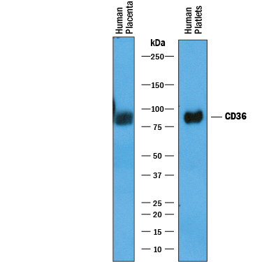
Detection of Human CD36/SR‑B3 by Western Blot. Western blot shows lysates of human placenta tissue and human platelets. PVDF membrane was probed with 1 µg/mL of Goat Anti-Human CD36/SR-B3 Antigen Affinity-purified Polyclonal Antibody (Catalog # AF1955) followed by HRP-conjugated Anti-Goat IgG Secondary Antibody (Catalog # HAF019). A specific band was detected for CD36/SR-B3 at approximately 85-90 kDa (as indicated). This experiment was conducted under reducing conditions and using Immunoblot Buffer Group 1.
 View Larger
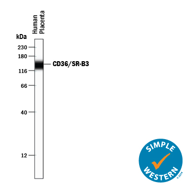
Detection of Human CD36/SR‑B3 by Simple WesternTM. Simple Western lane view shows lysates of human placenta tissue, loaded at 0.2 mg/mL. A specific band was detected for CD36/SR-B3 at approximately 140 kDa (as indicated) using 10 µg/mL of Goat Anti-Human CD36/SR-B3 Antigen Affinity-purified Polyclonal Antibody (Catalog # AF1955) followed by 1:50 dilution of HRP-conjugated Anti-Goat IgG Secondary Antibody (Catalog # HAF109). This experiment was conducted under reducing conditions and using the 12-230 kDa separation system.
 View Larger
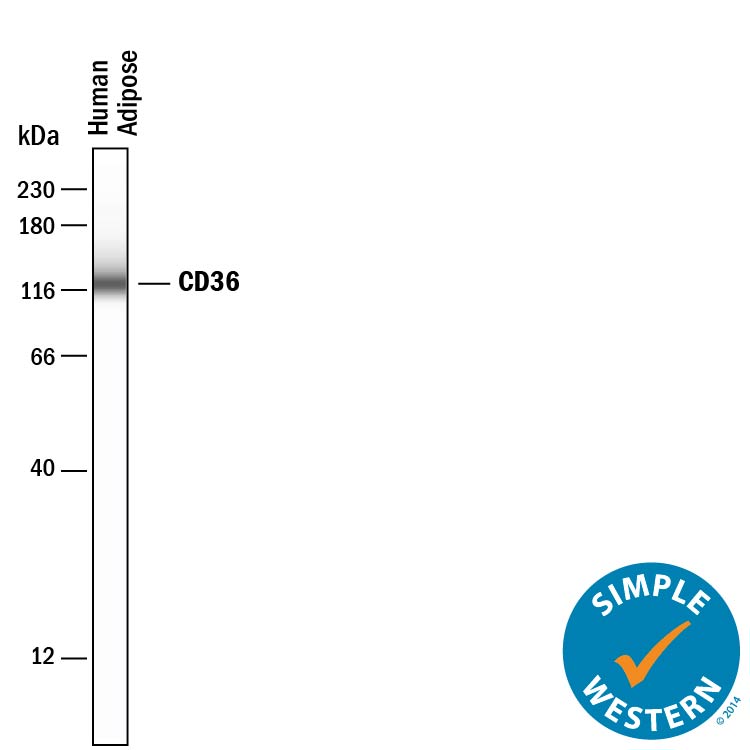
Detection of Human CD36/SR‑B3 by Simple WesternTM. Simple Western lane view shows lysates of human adipose tissue, loaded at 0.2 mg/mL. A specific band was detected for CD36/SR-B3 at approximately 125 kDa (as indicated) using 50 µg/mL of Goat Anti-Human CD36/SR-B3 Antigen Affinity-purified Polyclonal Antibody (Catalog # AF1955) followed by 1:50 dilution of HRP-conjugated Anti-Goat IgG Secondary Antibody (Catalog # HAF109). This experiment was conducted under reducing conditions and using the 12-230 kDa separation system.
Reconstitution Calculator
Preparation and Storage
- 12 months from date of receipt, -20 to -70 degreesC as supplied. 1 month, 2 to 8 degreesC under sterile conditions after reconstitution. 6 months, -20 to -70 degreesC under sterile conditions after reconstitution.
Background: CD36/SR-B3
CD36, alternatively known as platelet membrane glycoprotein IV (GPIV), GPIIIb, thrombospondin receptor, collagen receptor, fatty acid translocase (FAT), and scavenger receptor class B, member 3 (SR-B3), is an integral membrane glycoprotein that has multiple physiological functions (1). It is broadly expressed on a variety of cell types including microvascular endothelium, adipocytes, skeletal muscle, epithelial cells of the retina, breast, and intestine, smooth muscle cells, erythroid precursors, platelets, megakaryocytes, dendritic cells, monocytes/macrophages, and microglia (1, 2). As a member of the scavenger receptor family, CD36 is a multiligand pattern recognition receptor that interacts with a large number of structurally dissimilar ligands, including long chain fatty acid (LCFA), advanced glycation end products (AGE), thrombospondin-1, oxidized low-density lipoproteins (oxLDLs), high density lipoprotein (HDL), phosphatidylserine, apoptotic cells, beta -amyloid fibrils (fA beta ), collagens I and IV, and Plasmodium falciparum-infected erythrocytes (3). CD36 is required for the anti-angiogenic effects of thrombospondin-1 in the corneal neovascularization assay (4). It plays a role in lipid metabolism and has been identified as a fatty acid translocase necessary for the binding and transport of LCFA in cells and tissues (5). CD36 has been implicated in the clearance of apoptotic cells and cell debris and has also been shown to mediate the internalization and degradation of a variety of its ligands such as oxLDL, AGE and fA beta (3). Upon ligand binding, CD36 transduces signals that mediate a wide range of pro-inflammatory cellular responses (2). CD36 plays a significant role in the initiation and pathogenesis of chronic inflammatory diseases such as Alzheimer’s disease and atherosclerosis (2, 3). The human CD36 gene encodes a single-chain 472 amino acid residue protein containing both an N- and a C-terminal cytoplasmic tail and an extracellular loop.
- Febbraio, M. et al. (2001) J. Clin. Invest. 108:785.
- Khoury, J. et al. (2003) J. Exp. Med. 197:1657.
- Husemann, J. et al. (2002) Glia 40:195.
- Armstrong, L and P. Bornstein (2003) Matrix. Biol. 22:63.
- Febbraio M. et al. (1999) J. Biol. Chem. 274:19055.
Product Datasheets
Citations for Human CD36/SR-B3 Antibody
R&D Systems personnel manually curate a database that contains references using R&D Systems products. The data collected includes not only links to publications in PubMed, but also provides information about sample types, species, and experimental conditions.
8
Citations: Showing 1 - 8
Filter your results:
Filter by:
-
Microvascular insulin resistance associates with enhanced muscle glucose disposal in CD36 deficiency
Authors: Shibao, CA;Peche, VS;Williams, IM;Samovski, D;Pietka, TA;Abumrad, NN;Gamazon, E;Goldberg, I;Wasserman, D;Abumrad, NA;
medRxiv : the preprint server for health sciences
Species: Mouse
Sample Types: In Vivo
Applications: In vivo assay -
Human CEACAM1-LF regulates lipid storage in HepG2 cells via fatty acid transporter CD36
Authors: J Chean, CJ Chen, G Gugiu, P Wong, S Cha, H Li, T Nguyen, S Bhattichar, JE Shively
The Journal of Biological Chemistry, 2021-10-16;0(0):101311.
Species: Human
Sample Types: Cell Lysates
Applications: Immunoprecipitation, Western Blot -
DeSUMOylase SENP7-Mediated Epithelial Signaling Triggers Intestinal Inflammation via Expansion of Gamma-Delta T Cells
Authors: A Suhail, ZA Rizvi, P Mujagond, SA Ali, P Gaur, M Singh, V Ahuja, A Awasthi, CV Srikanth
Cell Rep, 2019-12-10;29(11):3522-3538.e7.
Species: Human
Sample Types: Cell Lysates
Applications: Western Blot -
Adipocyte-induced CD36 expression drives ovarian cancer progression and metastasis
Authors: A Ladanyi, A Mukherjee, HA Kenny, A Johnson, AK Mitra, S Sundaresan, KM Nieman, G Pascual, SA Benitah, A Montag, SD Yamada, NA Abumrad, E Lengyel
Oncogene, 2018-02-05;0(0):.
Species: Human
Sample Types: Cell Lysates
Applications: Western Blot -
Regulation of AMPK activation by CD36 links fatty acid uptake to beta-oxidation.
Authors: Samovski D, Sun J, Pietka T, Gross R, Eckel R, Su X, Stahl P, Abumrad N
Diabetes, 2014-08-25;64(2):353-9.
Species: Human
Sample Types: Cell Lysates
Applications: Western Blot -
A novel ELISA for measuring CD36 protein in human adipose tissue.
Authors: Allred CC, Krennmayr T, Koutsari C, Zhou L, Ali AH, Jensen MD
J. Lipid Res., 2010-11-28;52(2):408-15.
Species: Human
Sample Types: Cell Lysates, Tissue Homogenates
Applications: ELISA Development, Western Blot -
Preventive effects of heregulin-beta1 on macrophage foam cell formation and atherosclerosis.
Authors: Xu G, Watanabe T, Iso Y, Koba S, Sakai T, Nagashima M, Arita S, Hongo S, Ota H, Kobayashi Y, Miyazaki A, Hirano T
Circ. Res., 2009-07-30;105(5):500-10.
Species: Human
Sample Types: Cell Lysates
Applications: Western Blot -
Internal mammary artery smooth muscle cells resist migration and possess high antioxidant capacity.
Authors: Mahadevan VS, Campbell M, McKeown PP, Bayraktutan U
Cardiovasc. Res., 2006-06-27;72(1):60-8.
Species: Human
Sample Types: Cell Lysates
Applications: Western Blot
FAQs
No product specific FAQs exist for this product, however you may
View all Antibody FAQsReviews for Human CD36/SR-B3 Antibody
There are currently no reviews for this product. Be the first to review Human CD36/SR-B3 Antibody and earn rewards!
Have you used Human CD36/SR-B3 Antibody?
Submit a review and receive an Amazon gift card.
$25/€18/£15/$25CAN/¥75 Yuan/¥2500 Yen for a review with an image
$10/€7/£6/$10 CAD/¥70 Yuan/¥1110 Yen for a review without an image


