Human CD9 Antibody Summary
Ser112-Ile195
Accession # P21926
Applications
Please Note: Optimal dilutions should be determined by each laboratory for each application. General Protocols are available in the Technical Information section on our website.
Scientific Data
 View Larger
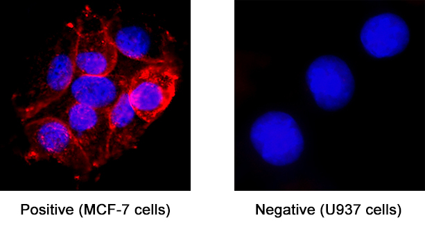
Detection of CD9 in MCF‑7 Human Breast Cancer Cell Line (Positive) and U937 Human Histiocytic Lymphoma Cell Line (Negative). CD9 was detected in immersion fixed MCF‑7 Human Breast Cancer Cell Line (Positive) and U937 Human Histiocytic Lymphoma Cell Line (Negative) using Mouse Anti-Human CD9 Monoclonal Antibody (Catalog # MAB10582) at 8 µg/mL for 3 hours at room temperature. Cells were stained using the NorthernLights™ 557-conjugated Anti-Mouse IgG Secondary Antibody (red; Catalog # NL007) and counterstained with DAPI (blue). Specific staining was localized to cell surface and cytoplasm. View our protocol for Fluorescent ICC Staining of Cells on Coverslips.
 View Larger
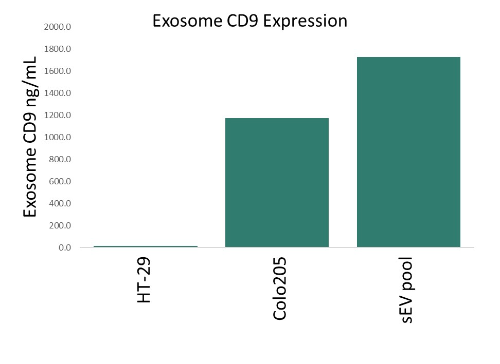
Detection of Human CD9 in exosomes with Anti-Human CD9 Monoclonal Antibody in ELISA Assay. The Human CD9 Antibody (Catalog # MAB10582) was conjugated with an affinity tag and incubated at 0.05ug/mL with culture media from HT29, COLO205 cell lines, and ultracentrifuge-enriched serum exosomes. HRP conjugated antibody was used as detection and incubated at 0.2ug/mL. The test was run on a microplate that was pre-coated with an anti-tag antibody
 View Larger
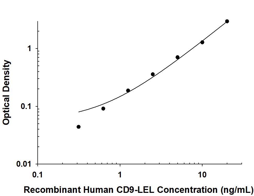
Detection of Human CD9 using Anti-Human CD9 Monoclonal Antibody in ELISA Assay. Recombinant Human CD9 protein (10015-CD) was serially diluted and incubated with Human CD9 Antibody (Catalog # MAB10582) that was conjugated with an affinity tag at 0.05ug/ml. HRP conjugated antibody was used as detection, incubated at 0.2ug/mL. The test was run on a microplate that was pre-coated with an anti-tag antibody.
Reconstitution Calculator
Preparation and Storage
- 12 months from date of receipt, -20 to -70 °C as supplied.
- 1 month, 2 to 8 °C under sterile conditions after reconstitution.
- 6 months, -20 to -70 °C under sterile conditions after reconstitution.
Background: CD9
CD9, also known as Tspan29, is a 24-27 kDa cell surface protein belonging to the tetraspanin family (1). Common to other tetraspanins, CD9 is composed of four transmembrane domains, short N- and C-terminal cytoplasmic domains, and two extracellular loops. The larger extracellular loop, referred to as the LEL or EC2, contains highly conserved CCG and PXSC motifs (2, 3). The LEL mediates noncovalent protein-protein interactions, allowing tetraspanins to associate with each other as well as signaling molecules, structural proteins, and G-protein coupled receptors (4-6). Human CD9 is expressed in multiple cell and tissue types and has been identified in diverse biological roles due to its involvement in the formation of tetraspanin-enriched microdomains (TEMs). TEMs are associated with numerous processes ranging from cell adhesion and fusion, membrane trafficking, and endocytosis to leukocyte adherence and motility (4-7). These tetraspanin-enriched microdomains (TEMs) are associated with a wide range of functions from cell adhesion and fusion, membrane trafficking and endocytosis, and eukocyte adherence and motility. The LEL of human CD9 shares 77% and 84% amino acid sequence identity with mouse and rat CD9, respectively. CD9 can form homodimers or interact with other proteins including CD117, CD29, CD46, CD49c, CD81, CD315, Tspan4, TGF-alpha, and HBEGF (1, 4, 8-13). Increased expression of CD9 has been shown to enhance transmembrane TGF-alpha -induced EGFR stimulation (1), and injection of human CD9 mRNA into CD9 knock-out mouse oocytes restored sperm-egg fusion (14). CD9-LEL may also be involved in the inhibition of multinucleated giant cell formation (3) as well as possess anti-adhesive effects against bacteria trying to invade mammalian cells (6, 15). CD9 interacts with integrins to regulate cell adhesion and motility (16-18). CD9 has been implicated in platelet activation and aggregation (17, 19). It may act as the terminal signal of myelination in the peripheral nervous system and can regulate the formation of paranodal junctions (20). Also, it has been suggested CD9 plays an important role both in the self-antigen and recall antigen-induced T cell activation (21).
- Shi, W. et al. (2000) J. Cell Biol. 148:591.
- Hemler, M. (2003) Annu Rev Cell Biol. 19:397.
- Hulme, R. et al. (2014) PLoS One 9:e116289.
- Stipp, C. et al. (2003) Trends Biochem Sci. 28:106.
- Barreiro, O. et al. (2005) Blood 105:2852.
- Ventress, J. et al. (2016) PLoS One 11:e0160387.
- Rubinstein, E. (2011) Biochem Soc Trans. 39:501.
- Anzai, N. et al. (2002) Blood 99:4413.
- Radford, K. et al. (1996) Biochem. Biophys. Res. Commun. 222:13.
- Lozahic, S. et al. (2000) Eur. J. Immunol. 30:900.
- Park, K. et al. (2000) Mol. Hum. Reprod. 6:252.
- Charrin, S. et al. (2001) J. Biol. Chem. 276:14329.
- Tachibana, I. et al. (1997) J. Biol. Chem. 272:29181.
- Zhu, G. et al. (2002) Development 129:1995.
- Green, L. et al. (2011) Infect Immun. 79:2241.
- Powner, D. et al. (2011) Biochem. Soc. Trans. 39:563.
- Detchokul, S. et al. (2014) British Journal of Pharmacology 171:5462.
- Reyes, R. et al. (2018) Front. Immunol. 9:863.
- Slupsky, J. et al. (1989) J Biol chem. 264:12289.
- Ishibashi, T. et al. (2004) J. Neuroscience 24:96.
- Kobayashi, H. et al. (2004) Clin Exp Immunol. 137:101.
Product Datasheets
FAQs
No product specific FAQs exist for this product, however you may
View all Antibody FAQsReviews for Human CD9 Antibody
There are currently no reviews for this product. Be the first to review Human CD9 Antibody and earn rewards!
Have you used Human CD9 Antibody?
Submit a review and receive an Amazon gift card.
$25/€18/£15/$25CAN/¥75 Yuan/¥2500 Yen for a review with an image
$10/€7/£6/$10 CAD/¥70 Yuan/¥1110 Yen for a review without an image

