Human CDO Antibody Summary
Asp26-Pro943 (Leu669Ile)
Accession # NP_058648
Applications
Please Note: Optimal dilutions should be determined by each laboratory for each application. General Protocols are available in the Technical Information section on our website.
Scientific Data
 View Larger
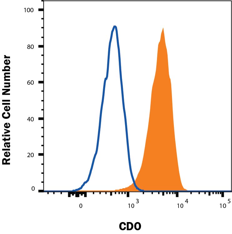
Detection of CDO in C2C12 Mouse Cell Line by Flow Cytometry. C2C12 mouse myoblast cell line was stained with Sheep Anti-Human CDO Antigen Affinity-purified Polyclonal Antibody (Catalog # AF4384, filled histogram) or control antibody (5-001-A, open histogram), followed by NorthernLights™ 557-conjugated Anti-Sheep IgG Secondary Antibody (NL010).
.") View Larger
View Larger
CDO in C2C12 Mouse Cell Line. CDO was detected in immersion fixed C2C12 mouse myoblast cell line using Sheep Anti-Human CDO Antigen Affinity-purified Polyclonal Antibody (Catalog # AF4384) at 10 µg/mL for 3 hours at room temperature. Cells were stained using the NorthernLights™ 557-conjugated Anti-Sheep IgG Secondary Antibody (red; NL010) and counterstained with DAPI (blue). Specific staining was localized to cell membranes. View our protocol for Fluorescent ICC Staining of Cells on Coverslips.
 View Larger
Detection of CDO in Jurkat cells by Flow Cytometry. Jurkat cells were stained with Sheep Anti-Human CDO Antigen Affinity-purified Polyclonal Antibody (Catalog # AF4384, filled histogram) or isotype control antibody (Catalog # 5-001-A, open histogram), followed by Phycoerythrin-conjugated Anti-Sheep IgG Secondary Antibody (Catalog # F0126). View our protocol for Staining Membrane-associated Proteins.
Reconstitution Calculator
Preparation and Storage
- 12 months from date of receipt, -20 to -70 °C as supplied.
- 1 month, 2 to 8 °C under sterile conditions after reconstitution.
- 6 months, -20 to -70 °C under sterile conditions after reconstitution.
Background: CDO
CDO (CAM-related/down‑regulated by oncogenes, also CDON; pronounced “kid-oh”) is a 190 kDa member of the Immunoglubulin (Ig) superfamily, Ig/Fibronectin (FN) type III repeat family of cell surface proteins (1). Human CDO is a type I transmembrane (TM) glycoprotein. It is synthesized as a 1287 amino acid (aa) precursor that contains a 25 aa signal sequence, a 938 aa extracellular domain (ECD), a 21 aa TM segment and a 303 aa cytoplasmic region (1, 2). The ECD contains five C2‑type Ig-like domains, followed by three FN type III repeats. The first FN repeat (aa 577‑673) is known to bind numerous cadherins, while the third (or juxtramembrane) FN type III repeat (aa 826‑923) binds SHH (3, 4). The intracellular region is believed to signal through various bHLH transcription factors (2). One alternate splice form is reported that shows a deletion of aa 1212‑1234 in the cytoplasmic tail. The ECD of human CDO is 85% aa identical to mouse CDO ECD. CDO is found on muscle precursor and neural progenitor cells of the embryo (5, 6). It likely promotes muscle differentiation, and contributes to axon guidance and neuronal patterning (2, 7, 8, 9). These effects may be mediated through two different receptor complexes. On muscle precursors, CDO apparently acts as both a coordinating and signaling subunit. Here, it integrates N- and M-cadherin, neogenin, netrin-3 and BOC into a cis-oriented receptor complex (2). While this complex has no identified ligand, intercellular cadherin interactions or netrin, may be enough to trigger CDO/cadherin/neogenin signaling. On axons, CDO may participate in a poorly‑defined receptor complex minimally composed of CDO, BOC and Gas1 that binds SHH, and interacts with PTCH1 (7, 8, 10).
- Kang, J.S. et al. (1997) J. Cell Biol. 138:203.
- Krauss, R.S. et al. (2005) J. Cell Sci. 118:2355.
- Yao, S. et al. (2006) Cell 125:343.
- Kang, J-S. et al. (2003) Proc. Natl. Acad. Sci. USA 100:3989.
- Kang, J-S. et al. (2002) EMBO J. 21:114.
- Zhang, W. et al. (2006) Mol. Cell. Biol. 26:3764.
- Okada, A. et al. (2006) Nature 444:369.
- Allen, B.L. et al. (2007) Genes Dev. 21:1244.
- Kang, J-S. et al. (2004) J. Cell. Biol. 167:493.
- Tenzen, T. et al. (2006) Dev. Cell 10:647.
Product Datasheets
Citation for Human CDO Antibody
R&D Systems personnel manually curate a database that contains references using R&D Systems products. The data collected includes not only links to publications in PubMed, but also provides information about sample types, species, and experimental conditions.
1 Citation: Showing 1 - 1
-
Cdon acts as a Hedgehog decoy receptor during proximal-distal patterning of the optic vesicle.
Authors: Cardozo M, Sanchez-Arrones L, Sandonis A, Sanchez-Camacho C, Gestri G, Wilson S, Guerrero I, Bovolenta P
Nat Commun, 2014-07-08;5(0):4272.
Species: Human
Sample Types: Cell Lysates
Applications: Western Blot
FAQs
No product specific FAQs exist for this product, however you may
View all Antibody FAQsReviews for Human CDO Antibody
There are currently no reviews for this product. Be the first to review Human CDO Antibody and earn rewards!
Have you used Human CDO Antibody?
Submit a review and receive an Amazon gift card.
$25/€18/£15/$25CAN/¥75 Yuan/¥2500 Yen for a review with an image
$10/€7/£6/$10 CAD/¥70 Yuan/¥1110 Yen for a review without an image

