Human Cystatin C Antibody Summary
Applications
Please Note: Optimal dilutions should be determined by each laboratory for each application. General Protocols are available in the Technical Information section on our website.
Scientific Data
 View Larger
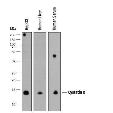
Detection of Human Cystatin C by Western Blot. Western blot shows lysates of HepG2 human hepatocellular carcinoma cell line, human liver tissue, and human serum. PVDF membrane was probed with 0.25 µg/mL of Goat Anti-Human Cystatin C Antigen Affinity-purified Polyclonal Antibody (Catalog # AF1196) followed by HRP-conjugated Anti-Goat IgG Secondary Antibody (Catalog # HAF109). A specific band was detected for Cystatin C at approximately 14 kDa (as indicated). This experiment was conducted under reducing conditions and using Immunoblot Buffer Group 1.
 View Larger
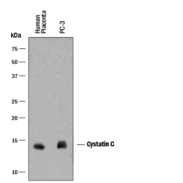
Detection of Human Cystatin C by Western Blot. Western blot shows lysates of human placenta tissue and PC-3 human prostate cancer cell line. PVDF membrane was probed with 2 µg/mL of Goat Anti-Human Cystatin C Antigen Affinity-purified Polyclonal Antibody (Catalog # AF1196) followed by HRP-conjugated Anti-Goat IgG Secondary Antibody (Catalog # HAF017). A specific band was detected for Cystatin C at approximately 14 kDa (as indicated). This experiment was conducted under reducing conditions and using Immunoblot Buffer Group 1.
 View Larger
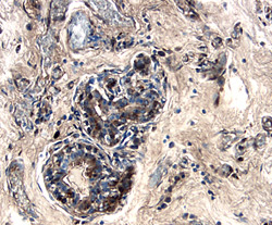
Cystatin C in Human Breast. Cystatin C was detected in immersion fixed paraffin-embedded sections of human breast using 5 µg/mL Goat Anti-Human Cystatin C Antigen Affinity-purified Polyclonal Antibody (Catalog # AF1196) overnight at 4 °C. Tissue was stained with the Anti-Goat HRP-DAB Cell & Tissue Staining Kit (brown; Catalog # CTS008) and counterstained with hematoxylin (blue). Specific labeling was localized to the cytoplasm in cells of terminal ductules. View our protocol for Chromogenic IHC Staining of Paraffin-embedded Tissue Sections.
 View Larger
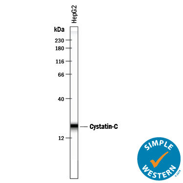
Detection of Human Cystatin C by Simple WesternTM. Simple Western lane view shows lysates of HepG2 human hepatocellular carcinoma cell line, loaded at 0.2 mg/mL. A specific band was detected for Cystatin C at approximately 20 kDa (as indicated) using 10 µg/mL of Goat Anti-Human Cystatin C Antigen Affinity-purified Polyclonal Antibody (Catalog # AF1196). This experiment was conducted under reducing conditions and using the 12-230 kDa separation system.
Reconstitution Calculator
Preparation and Storage
- 12 months from date of receipt, -20 to -70 degreesC as supplied. 1 month, 2 to 8 degreesC under sterile conditions after reconstitution. 6 months, -20 to -70 degreesC under sterile conditions after reconstitution.
Background: Cystatin C
Cystatin C is a member of family 2 of the Cystatin superfamily (1). It is involved in processes such as tumor invasion and metastasis, inflammation and some neurological diseases. It inhibits many cysteine proteases such as papain and cathepsins B, H, K, L, and S (2, 3). It is ubiquitous in human tissues and body fluids. A point mutation in the gene coding for the 120 amino acid mature Cystatin C causes a hereditary form of amyloid angiopathy in which the protein variant (Leu68 to Gln) is deposited in the cerebral arteries, leading to fatal cerebral hemorrhage (4). Cystatin C may have additional clinical applications. For example, it is a good marker for glomerular filtration rate (5).
- Reed, C.H. (2000) British J. Biomed. Sci. 57:323.
- Janowski, R. et al. (2001) Nat. Struct. Biol. 8:316.
- Abrahamson, M. (1994) Methods Enzymol. 244:685.
- Abrahamson, M. et al. (1992) Hum. Genet. 89:377.
- Laterza, O.F. et al. (2002) Clin. Chem. 48:699.
Product Datasheets
Citations for Human Cystatin C Antibody
R&D Systems personnel manually curate a database that contains references using R&D Systems products. The data collected includes not only links to publications in PubMed, but also provides information about sample types, species, and experimental conditions.
3
Citations: Showing 1 - 3
Filter your results:
Filter by:
-
Nanoparticle Detection of Urinary Markers for Point-of-Care Diagnosis of Kidney Injury.
Authors: Chung H, Pellegrini K, Chung J, Wanigasuriya K, Jayawardene I, Lee K, Lee H, Vaidya V, Weissleder R
PLoS ONE, 2015-07-17;10(7):e0133417.
Species: Human
Sample Types: Urine
Applications: ELISA Development -
Cystatin E/M suppresses legumain activity and invasion of human melanoma.
Authors: Briggs JJ, Haugen MH, Johansen HT, Riker AI, Abrahamson M, Fodstad O, Maelandsmo GM, Solberg R
BMC Cancer, 2010-01-15;10(1):17.
Species: Human
Sample Types: Cell Lysates
Applications: Western Blot -
Proteomic analysis of factors released from p21-overexpressing tumour cells.
Authors: Currid CA, O&apos;Connor DP, Chang BD, Gebus C, Harris N, Dawson KA, Dunn MJ, Pennington SR, Roninson IB, Gallagher WM
Proteomics, 2006-07-01;6(13):3739-53.
Species: Human
Sample Types: Cell Culture Supernates
Applications: Array Development
FAQs
No product specific FAQs exist for this product, however you may
View all Antibody FAQsReviews for Human Cystatin C Antibody
Average Rating: 4.5 (Based on 2 Reviews)
Have you used Human Cystatin C Antibody?
Submit a review and receive an Amazon gift card.
$25/€18/£15/$25CAN/¥75 Yuan/¥2500 Yen for a review with an image
$10/€7/£6/$10 CAD/¥70 Yuan/¥1110 Yen for a review without an image
Filter by:
