Human Cytokeratin 19 Antibody Summary
Thr2-Leu400
Accession # P08727
Applications
Please Note: Optimal dilutions should be determined by each laboratory for each application. General Protocols are available in the Technical Information section on our website.
Scientific Data
 View Larger
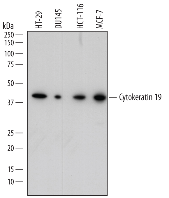
Detection of Human Cytokeratin 19 by Western Blot. Western blot shows lysates of HT-29 human colon adenocarcinoma cell line, DU145 human prostate carcinoma cell line, HCT-116 human colorectal carcinoma cell line, and MCF-7 human breast cancer cell line. PVDF membrane was probed with 0.1 µg/mL of Sheep Anti-Human Cytokeratin 19 Antigen Affinity-purified Polyclonal Antibody (Catalog # AF3506) followed by HRP-conjugated Anti-Sheep IgG Secondary Antibody (Catalog # HAF016). A specific band was detected for Cytokeratin 19 at approximately 40 kDa (as indicated). This experiment was conducted under reducing conditions and using Immunoblot Buffer Group 1.
 View Larger
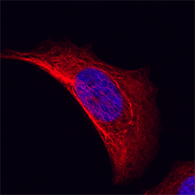
Cytokeratin 19 in MCF‑7 Human Cell Line. Cytokeratin 19 was detected in immersion fixed MCF-7 human breast cancer cell line using Sheep Anti-Human Cytokeratin 19 Antigen Affinity-purified Polyclonal Antibody (Catalog # AF3506) at 10 µg/mL for 3 hours at room temperature. Cells were stained using the NorthernLights™ 557-conjugated Anti-Sheep IgG Secondary Antibody (red; Catalog # NL010) and counterstained with DAPI (blue). Specific staining was localized to cytoplasm. View our protocol for Fluorescent ICC Staining of Cells on Coverslips.
 View Larger
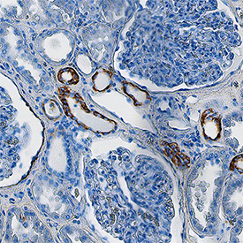
Cytokeratin 19 in Human Kidney. Cytokeratin 19 was detected in immersion fixed paraffin-embedded sections of human kidney using Sheep Anti-Human Cytokeratin 19 Antigen Affinity-purified Polyclonal Antibody (Catalog # AF3506) at 3 µg/mL overnight at 4 °C. Tissue was stained using the Anti-Sheep HRP-DAB Cell & Tissue Staining Kit (brown; Catalog # CTS019) and counterstained with hematoxylin (blue). Specific staining was localized to cytoplasm in convoluted tubules. View our protocol for Chromogenic IHC Staining of Paraffin-embedded Tissue Sections.
 View Larger
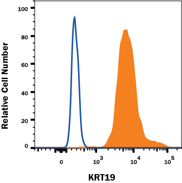
Detection of KRT19 in Human MCF-7 cell line by Flow Cytometry. MCF-7 human breast cancer cell line was stained with Sheep Anti-Human KRT19 Affinity Purified Polyclonal Antibody (Catalog # AF3506, filled histogram) or control antibody (Catalog # 5-001-A, open histogram) followed by Anti-Sheep IgG PE-conjugated secondary antibody (Catalog # F0126). To facilitate intracellular staining, cells were fixed and permeabilized with FlowX FoxP3 Fixation & Permeabilization Buffer Kit (Catalog # FC012). View our protocol for Staining Membrane-associated Proteins.
 View Larger
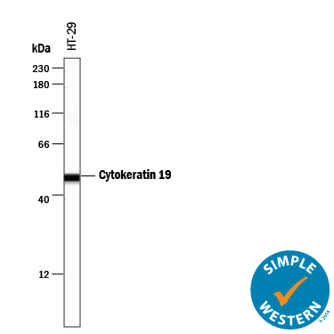
Detection of Human Cytokeratin 19 by Simple WesternTM. Simple Western lane view shows lysates of HT-29 human colon adenocarcinoma cell line, loaded at 0.2 mg/mL. A specific band was detected for Cytokeratin 19 at approximately 50 kDa (as indicated) using 1 µg/mL of Sheep Anti-Human Cytokeratin 19 Antigen Affinity-purified Polyclonal Antibody (Catalog # AF3506) followed by 1:50 dilution of HRP-conjugated Anti-Sheep IgG Secondary Antibody (Catalog # HAF016). This experiment was conducted under reducing conditions and using the 12-230 kDa separation system.
Reconstitution Calculator
Preparation and Storage
- 12 months from date of receipt, -20 to -70 °C as supplied.
- 1 month, 2 to 8 °C under sterile conditions after reconstitution.
- 6 months, -20 to -70 °C under sterile conditions after reconstitution.
Background: Cytokeratin 19
Cytokeratin 19 (Keratin, type I cytoskeletal 19; also KRT-19, CK19 and Keratin-19) is a 40-45 kDa, acidic Class I keratin member of the intermediate filament family of proteins. Individual keratins are always expressed in tandem with a second keratin, and these are found in all epithelial cells. The class I KRT-19 heterodimerizes/polymerizes with 50-52 kDa class II KRT-8 (plus KRT-5 and -7) to form 8-10 nm filaments in epidermal stem cells, secretory gland (sweat; mammary; bile duct) simple epithelium, and neuroendocrine epidermal Merkel cells. It may represent a viable marker for skin stem cells. In skin, Cytokeratin 19 forms filaments in the fetal epithelium, and then progressively decreases with age, being virtually absent by age 17. Human Cytokeratin 19 is 400 amino acids (aa) in length. It contains an N-terminal "head" region (aa 1-79) and a subsequent "rod" region (aa 80-387), but is absent a typical C-terminal tail region. Cytokeratin 19 possesses at least 5 utilized phosphorylation sites plus one acetylated Lys residue. Based on other keratins, and the presence of an Asp at position 238, there may be caspase cleavage-generated isoforms. Full length human Cytokeratin 19 (aa 2-400) shares 82% aa sequence identity with mouse Cytokeratin 19.
Product Datasheets
Citation for Human Cytokeratin 19 Antibody
R&D Systems personnel manually curate a database that contains references using R&D Systems products. The data collected includes not only links to publications in PubMed, but also provides information about sample types, species, and experimental conditions.
1 Citation: Showing 1 - 1
-
Murine- and Human-Derived Autologous Organoid/Immune Cell Co-Cultures as Pre-Clinical Models of Pancreatic Ductal Adenocarcinoma
Authors: L Holokai, J Chakrabart, J Lundy, D Croagh, P Adhikary, SS Richards, C Woodson, N Steele, R Kuester, A Scott, M Khreiss, T Frankel, J Merchant, BJ Jenkins, J Wang, RT Shroff, SA Ahmad, Y Zavros
Cancers, 2020-12-17;12(12):.
Species: Human
Sample Types: Organoids
Applications: ICC
FAQs
No product specific FAQs exist for this product, however you may
View all Antibody FAQsReviews for Human Cytokeratin 19 Antibody
There are currently no reviews for this product. Be the first to review Human Cytokeratin 19 Antibody and earn rewards!
Have you used Human Cytokeratin 19 Antibody?
Submit a review and receive an Amazon gift card.
$25/€18/£15/$25CAN/¥75 Yuan/¥2500 Yen for a review with an image
$10/€7/£6/$10 CAD/¥70 Yuan/¥1110 Yen for a review without an image


