Human DC-SIGN/CD209 Antibody Summary
Lys62-Ala404
Accession # Q9NNX6
Applications
Please Note: Optimal dilutions should be determined by each laboratory for each application. General Protocols are available in the Technical Information section on our website.
Scientific Data
 View Larger
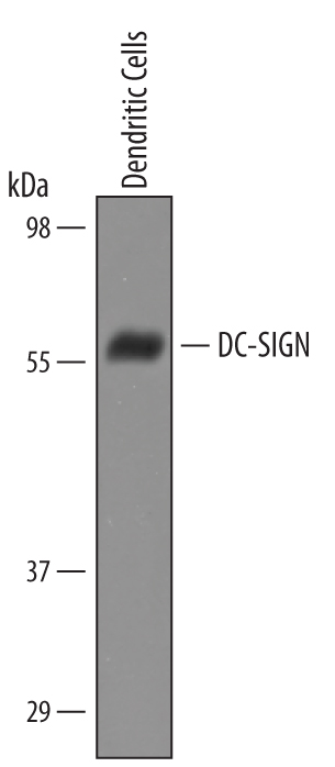
Detection of Human DC‑SIGN/CD209 by Western Blot. Western blot shows lysates of human dendritic cells. PVDF Membrane was probed with 1 µg/mL of Sheep Anti-Human DC-SIGN/CD209 Antigen Affinity-purified Polyclonal Antibody (Catalog # AF161) followed by HRP-conjugated Anti-Goat IgG Secondary Antibody (Catalog # HAF019). A specific band was detected for DC-SIGN/CD209 at approximately 50 kDa (as indicated). This experiment was conducted under reducing conditions and using Immunoblot Buffer Group 8.
Reconstitution Calculator
Preparation and Storage
- 12 months from date of receipt, -20 to -70 °C as supplied.
- 1 month, 2 to 8 °C under sterile conditions after reconstitution.
- 6 months, -20 to -70 °C under sterile conditions after reconstitution.
Background: DC-SIGN/CD209
Human DC-SIGN (dendritic cell-specific ICAM-3 grabbing nonintegrin; also CD209) is a member of the chromosome 19 C-type lectin family that includes DC-SIGN, DC-SIGN-related protein, CD23 and LSECtin (1). DC-SIGN was initially reported to be a 46 kDa, 404 amino acid (aa) type II transmembrane protein that contained a 40 aa cytoplasmic N-terminus, a 21 aa transmembrane segment, and a 343 aa extracellular C-terminus (2). The extracellular region contains a distal, 115 aa Ca++‑dependent carbohydrate-binding lectin domain and a membrane-proximal linker segment that is composed of seven 23 aa repeats (2, 3). The lectin domain is believed to preferably bind mannose, either within the context of ICAM-3 (on T cells) or ICAM-2 (on endothelial cells) (2, 4, 5). DC-SIGN expression appears to be limited to dendritic cells (DC) and macrophages (6), and DC interaction with the ICAMs both aids DC cell trafficking and immunological synapse formation (7). Since the original report on DC-SIGN, multiple splice forms have been discovered, generating both membrane-bound and soluble forms (3). There are eight type A isoforms, all of which begin with the same 15 aa of exon 1a. Four contain the transmembrane region of exon II, and four do not (i.e., are soluble). Among these eight type A isoforms, only three retain the entire 343 aa found in the full length form described in reference #2 (the full length form is referred to as type I mDC-SIGN1A) (3). Five additional isoforms utilize an alternate start site, and these are referred to as type B isoforms. These all show a 35 aa cytoplasmic domain. One also has a transmembrane segment; four do not. Two of the five contain full, unspliced extracellular regions (3). All of this suggests enormous complexity in DC-SIGN biology. DC-SIGN is not well conserved across species. Human and mouse show little overall aa identity. In the lectin domain, however, human is 68% aa identical to mouse (8). Human to rhesus monkey, there is 91% aa identity over the entire extracellular region (8).
- Liu, W. et al. (2004) J. Biol. Chem. 279:18748.
- Curtis, B.M. et al. (1992) Proc. Natl. Acad. Sci. USA 89:8356.
- Mummidi, S. et al. (2001) J. Biol. Chem. 276:33196.
- Su, S.V. et al. (2004) J. Biol. Chem. 279:19122.
- Cambi, A. et al. (2005) Cell. Microbiol. 7:481.
- Serrano-Gomez, D. et al. (2004) J. Immunol. 173:5635.
- Geijtenbeek, T.B.H. and Y. van Kooyk (2003) Curr. Top. Microbiol. Immunol. 276:32.
- Baribaud, F. et al. (2001) J. Virol. 75:10281.
Product Datasheets
FAQs
No product specific FAQs exist for this product, however you may
View all Antibody FAQsReviews for Human DC-SIGN/CD209 Antibody
There are currently no reviews for this product. Be the first to review Human DC-SIGN/CD209 Antibody and earn rewards!
Have you used Human DC-SIGN/CD209 Antibody?
Submit a review and receive an Amazon gift card.
$25/€18/£15/$25CAN/¥75 Yuan/¥2500 Yen for a review with an image
$10/€7/£6/$10 CAD/¥70 Yuan/¥1110 Yen for a review without an image


