Human DC-SIGN/CD209 PE-conjugated Antibody
Human DC-SIGN/CD209 PE-conjugated Antibody Summary
Applications
Please Note: Optimal dilutions should be determined by each laboratory for each application. General Protocols are available in the Technical Information section on our website.
Scientific Data
 View Larger
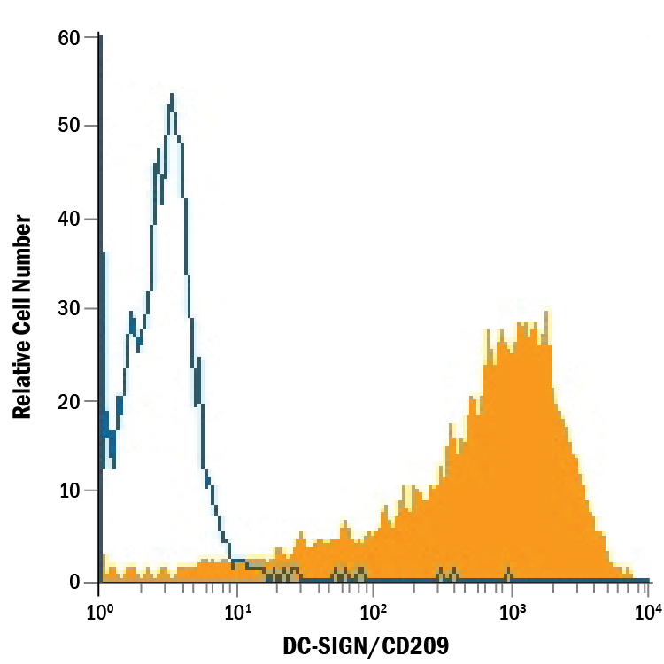
Detection of DC‑SIGN/CD209 in NIH‑3T3 Mouse Cell Line Transfected with Human DC-SIGN/CD209 by Flow Cytometry. NIH-3T3 mouse embryonic fibroblast cell line transfected with human DC-SIGN/CD209 was stained with Mouse Anti-Human DC-SIGN/CD209 PE-conjugated Monoclonal Antibody (Catalog # FAB161P, filled histogram) or isotype control antibody (Catalog # IC0041P, open histogram). View our protocol for Staining Membrane-associated Proteins.
Reconstitution Calculator
Preparation and Storage
- 12 months from date of receipt, 2 to 8 °C as supplied.
Background: DC-SIGN/CD209
Human DC-SIGN (dendritic cell-specific ICAM-3 grabbing nonintegrin; also known as CD209) is a member of the chromosome 19 C-type lectin family that includes DC-SIGN, DC-SIGN-related protein, CD23 and LSECtin (1). DC-SIGN was initially reported to be a 46 kDa, 404 amino acid (aa) type II transmembrane protein that contained a 40 aa cytoplasmic N-terminus, a 21 aa transmembrane segment, and a 343 aa extracellular C-terminus (2). The extracellular region contains a distal, 115 aa Ca++-dependent carbohydrate-binding lectin domain and a membrane-proximal linker segment that is composed of seven 23 aa repeats (2, 3). The lectin domain is believed to preferably bind mannose, either within the context of ICAM-3 (on T cells) or ICAM-2 (on endothelial cells) (2, 4, 5). DC-SIGN expression appears to be limited to dendritic cells (DC) and macrophages (6), and DC interaction with the ICAMs both aids DC cell trafficking and immunological synapse formation (7). Since the original report on DC-SIGN, multiple splice forms have been discovered, generating both membrane-bound and soluble forms (3). There are eight type A isoforms, all of which begin with the same 15 aa of exon 1a. Four contain the transmembrane region of exon II, and four do not (i.e., are soluble). Among these eight type A isoforms, only three retain the entire 343 aa found in the full length form described in reference #2 (the full length form is referred to as type I mDC-SIGN1A) (3). Five additional isoforms utilize an alternate start site, and these are referred to as type B isoforms. These all show a 35 aa cytoplasmic domain. One also has a transmembrane segment; four do not. Two of the five contain full, unspliced extracellular regions (3). All of this suggests enormous complexity in DC-SIGN biology. DC-SIGN is not well conserved across species. Human and mouse show little overall aa identity. In the lectin domain, however, human DC-SIGN shares 68% aa identity with mouse DC-SIGN (8). Human and rhesus monkey DC-SIGN share 91% aa identity over the entire extracellular region (8). A detailed description of the additional properties of this monoclonal antibody (MAB161) have been published (9, 10).
- Liu, W. et al. (2004) J. Biol. Chem. 279:18748.
- Curtis, B.M. et al. (1992) Proc. Natl. Acad. Sci. USA 89:8356.
- Mummidi, S. et al. (2001) J. Biol. Chem. 276:33196.
- Su, S.V. et al. (2004) J. Biol. Chem. 279:19122.
- Cambi, A. et al. (2005) Cell. Microbiol. 7:481.
- Serrano-Gomez, D. et al. (2004) J. Immunol. 173:5635.
- Geijtenbeek, T.B.H. and Y. van Kooyk (2003) Curr. Top. Microbiol. Immunol. 276:32.
- Baribaud, F. et al. (2001) J. Virol. 75:10281.
- Wu, L. et al. (2002) J. Virol. 76:5905.
- Baribaud, F. et al. (2002) J. Virol.76:9135.
Product Datasheets
Citations for Human DC-SIGN/CD209 PE-conjugated Antibody
R&D Systems personnel manually curate a database that contains references using R&D Systems products. The data collected includes not only links to publications in PubMed, but also provides information about sample types, species, and experimental conditions.
11
Citations: Showing 1 - 10
Filter your results:
Filter by:
-
Elevated glycolytic metabolism of monocytes limits the generation of HIF1A-driven migratory dendritic cells in tuberculosis
Authors: Maio, M;Barros, J;Joly, M;Vahlas, Z;Marín Franco, JL;Genoula, M;Monard, SC;Vecchione, MB;Fuentes, F;Gonzalez Polo, V;Quiroga, MF;Vermeulen, M;Vu Manh, TP;Argüello, RJ;Inwentarz, S;Musella, R;Ciallella, L;González Montaner, P;Palmero, D;Lugo Villarino, G;Sasiain, MDC;Neyrolles, O;Vérollet, C;Balboa, L;
eLife
Species: Human
Sample Types: Whole Cells
Applications: Immunocytochemistry -
Early Colorectal Responses to HIV-1 and Modulation by Antiretroviral Drugs
Authors: C Herrera, MD McRaven, KG Laing, J Dennis, TJ Hope, RJ Shattock
Vaccines, 2021-03-08;9(3):.
Species: Human
Sample Types: Whole Tissue
Applications: Flow Cytometry -
Fatty acid oxidation of alternatively activated macrophages prevents foam cell formation, but Mycobacterium tuberculosis counteracts this process via HIF-1&alpha activation
Authors: M Genoula, JL Marín Fran, M Maio, B Dolotowicz, M Ferreyra, MA Milillo, R Mascarau, EJ Moraña, D Palmero, M Matteo, F Fuentes, B López, P Barrionuev, O Neyrolles, C Cougoule, G Lugo-Villa, C Vérollet, MDC Sasiain, L Balboa
PLoS Pathog, 2020-10-01;16(10):e1008929.
Species: Human
Sample Types: Whole Cells
Applications: Flow Cytometry -
A Comprehensive Map of the Monocyte-Derived Dendritic Cell Transcriptional Network Engaged upon Innate Sensing of HIV
Authors: JS Johnson, N De Veaux, AW Rives, X Lahaye, SY Lucas, BP Perot, M Luka, V Garcia-Par, LM Amon, A Watters, G Abdessalem, A Aderem, N Manel, DR Littman, R Bonneau, MM Ménager
Cell Rep, 2020-01-21;30(3):914-931.e9.
Species: Human
Sample Types: Whole Cells
-
Reshaping of the Dendritic Cell Chromatin Landscape and Interferon Pathways during HIV Infection
Authors: JS Johnson, SY Lucas, LM Amon, S Skelton, R Nazitto, S Carbonetti, DN Sather, DR Littman, A Aderem
Cell Host Microbe, 2018-03-14;23(3):366-381.e9.
Species: Human
Sample Types: Whole Cells
Applications: Flow Cytometry -
NK cell heparanase controls tumor invasion and immune surveillance
Authors: EM Putz, AJ Mayfosh, K Kos, DS Barkauskas, K Nakamura, L Town, KJ Goodall, DY Yee, IK Poon, N Baschuk, F Souza-Fons, MD Hulett, MJ Smyth
J. Clin. Invest., 2017-06-05;0(0):.
Species: Human
Sample Types: Whole Cells
Applications: Flow Cytometry -
Dendritic cells from the human female reproductive tract rapidly capture and respond to HIV
Mucosal Immunol, 2016-08-31;0(0):.
Species: Human
Sample Types: Whole Cells
Applications: Flow Cytometry -
HCV RNA Activates APCs via TLR7/TLR8 While Virus Selectively Stimulates Macrophages Without Inducing Antiviral Responses
Sci Rep, 2016-07-07;6(0):29447.
Species: Human
Sample Types: Whole Cells
Applications: Flow Cytometry -
Dendritic cell-induced activation of latent HIV-1 provirus in actively proliferating primary T lymphocytes.
Authors: van der Sluis R, van Montfort T, Pollakis G, Sanders R, Speijer D, Berkhout B, Jeeninga R
PLoS Pathog, 2013-03-21;9(3):e1003259.
Species: Human
Sample Types: Whole Cells
Applications: Flow Cytometry -
In vitro characterization of primary SIVsmm isolates belonging to different lineages. In vitro growth on rhesus macaque cells is not predictive for in vivo replication in rhesus macaques.
Authors: Gautam R, Carter AC, Katz N, Butler IF, Barnes M, Hasegawa A, Ratterree M, Silvestri G, Marx PA, Hirsch VM, Pandrea I, Apetrei C
Virology, 2007-02-15;362(2):257-70.
Species: Primate - Macaca mulatta (Rhesus Macaque)
Sample Types: Whole Cells
Applications: Flow Cytometry -
DC-SIGN is the major Mycobacterium tuberculosis receptor on human dendritic cells.
Authors: Tailleux L, Schwartz O, Herrmann JL, Pivert E, Jackson M, Amara A, Legres L, Dreher D, Nicod LP, Gluckman JC, Lagrange PH, Gicquel B, Neyrolles O
J. Exp. Med., 2003-01-06;197(1):121-7.
Species: Human
Sample Types: Whole Cells
Applications: Flow Cytometry
FAQs
No product specific FAQs exist for this product, however you may
View all Antibody FAQsReviews for Human DC-SIGN/CD209 PE-conjugated Antibody
There are currently no reviews for this product. Be the first to review Human DC-SIGN/CD209 PE-conjugated Antibody and earn rewards!
Have you used Human DC-SIGN/CD209 PE-conjugated Antibody?
Submit a review and receive an Amazon gift card.
$25/€18/£15/$25CAN/¥75 Yuan/¥2500 Yen for a review with an image
$10/€7/£6/$10 CAD/¥70 Yuan/¥1110 Yen for a review without an image


