Human EGFR PE-conjugated Antibody Summary
Applications
Please Note: Optimal dilutions should be determined by each laboratory for each application. General Protocols are available in the Technical Information section on our website.
Scientific Data
 View Larger
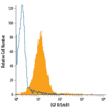
Detection of EGF R/ErbB1 in A431 human epithelial carcinoma cell line by Flow Cytometry. A431 human epithelial carcinoma cell line was stained with Rat Anti-Human EGF R/ErbB1 PE-conjugated Monoclonal Antibody (Catalog # FAB10951P, filled histogram) or isotype control antibody (Catalog # IC006P, open histogram). View our protocol for Staining Membrane-associated Proteins.
Reconstitution Calculator
Preparation and Storage
- 12 months from date of receipt, 2 to 8 degreesC as supplied.
Background: EGFR
Epidermal Growth Factor Receptor (EGF R), also named erythroblastic leukemia viral oncogene homolog 1 (ErbB1), is a member of the type I receptor tyrosine kinase superfamily. The epidermal growth factor receptor (EGF R) subfamily of receptor tyrosine kinases comprises four members: EGF R (also known as HER1, ErbB1or ErbB), ErbB2 (Neu, HER2), ErbB3 (HER3), and ErbB4 (HER4). All family members are type I transmembrane glycoproteins that have an extracellular domain with two ligand binding cysteine rich domains, separated by a spacer region, and a cytoplasmic domain with a membrane proximal tyrosine kinase domain and a C-terminal tail with multiple tyrosine autophosphorylation sites. The human EGF R geneencodes a 1210 amino acid (aa) residue precursor with a 24 aa putative signal peptide, a 621 aa extracellular domain, a 23 aa transmembrane domain, and a 542 aa cytoplasmic domain. EGF R has been shown to bind a subset of the EGF family ligands, including EGF, amphiregulin, TGF alpha, betacellulin, epiregulin, heparin-binding EGF and neuregulin-2 alpha , in the absence of a coreceptor. Ligand binding induces EGF R homodimerization as well as heterodimerization with ErbB2, resulting in kinase activation, tyrosine phosphorylation and cell signaling. EGF R can also be recruited to form heterodimers with ligand-activated ErbB3 or ErbB4. EGF R signaling has been shown to regulate multiple biological functions including cell proliferation, differentiation, motility and apoptosis. In addition, EGF R signaling has also been shown to play a role in carcinogenesis (1 - 3).
- Daly, R.J. (1999) Growth Factors, 16:255.
- Schlessinger, J. (2000) Cell. 103:211.
- Maihle, N.J. et al. (2002) Cancer Treat. Res. 107:247.
Product Datasheets
Citations for Human EGFR PE-conjugated Antibody
R&D Systems personnel manually curate a database that contains references using R&D Systems products. The data collected includes not only links to publications in PubMed, but also provides information about sample types, species, and experimental conditions.
6
Citations: Showing 1 - 6
Filter your results:
Filter by:
-
Decreased expression of ErbB2 on left ventricular epicardial cells in patients with diabetes mellitus
Authors: JT de Kay, J Carver, B Shevenell, AM Kosta, S Tsibulniko, E Certo, DB Sawyer, S Ryzhov, MP Robich
Cellular Signalling, 2022-05-21;96(0):110360.
Species: Human
Sample Types: Whole Cells
Applications: Flow Cytometry -
Infectious titer determination of lentiviral vectors using a temporal immunological real-time imaging approach
Authors: JJ Labisch, GP Wiese, K Barnes, F Bollmann, K Pflanz
PLoS ONE, 2021-07-15;16(7):e0254739.
Species: Human
Sample Types: Whole Cells
Applications: Flow Cytometry -
A new simplified clarification approach for lentiviral vectors using diatomaceous earth improves throughput and safe handling
Authors: JJ Labisch, F Bollmann, MW Wolff, K Pflanz
Journal of biotechnology, 2020-12-07;326(0):11-20.
Species: Human
Sample Types: Whole Cells
Applications: ICC -
Cetuximab-induced natural killer cell cytotoxicity in head and neck squamous cell carcinoma cell lines: investigation of the role of cetuximab sensitivity and HPV status
Authors: H Baysal, I De Pauw, H Zaryouh, J De Waele, M Peeters, P Pauwels, JB Vermorken, E Smits, F Lardon, J Jacobs, A Wouters
Br. J. Cancer, 2020-06-16;0(0):.
Species: Human
Sample Types: Whole Cells
Applications: Flow Cytometry -
Simultaneous targeting of EGFR, HER2 and HER4 by afatinib overcomes intrinsic and acquired cetuximab resistance in head and neck squamous cell carcinoma cell lines
Authors: I De Pauw, F Lardon, J Van den Bo, H Baysal, E Fransen, V Deschoolme, P Pauwels, M Peeters, JB Vermorken, A Wouters
Mol Oncol, 2018-05-01;0(0):.
Species: Human
Sample Types: Whole Cells
Applications: Flow Cytometry -
Glioblastoma stem-like cell lines with either maintenance or loss of high-level EGFR amplification, generated via modulation of ligand concentration.
Authors: Schulte A, Gunther HS, Martens T, Zapf S, Riethdorf S, Wulfing C, Stoupiec M, Westphal M, Lamszus K
Clin. Cancer Res., 2012-02-07;18(7):1901-13.
Species: Human
Sample Types: Whole Cells
Applications: Flow Cytometry
FAQs
No product specific FAQs exist for this product, however you may
View all Antibody FAQsReviews for Human EGFR PE-conjugated Antibody
There are currently no reviews for this product. Be the first to review Human EGFR PE-conjugated Antibody and earn rewards!
Have you used Human EGFR PE-conjugated Antibody?
Submit a review and receive an Amazon gift card.
$25/€18/£15/$25CAN/¥75 Yuan/¥2500 Yen for a review with an image
$10/€7/£6/$10 CAD/¥70 Yuan/¥1110 Yen for a review without an image


