Human EMMPRIN/CD147 Antibody Summary
Glu138-Ala323
Accession # P35613.2
Applications
Please Note: Optimal dilutions should be determined by each laboratory for each application. General Protocols are available in the Technical Information section on our website.
Scientific Data
 View Larger
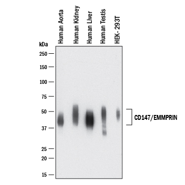
Detection of Human EMMPRIN/CD147 by Western Blot. Western blot shows lysates of Human heart (aorta), human kidney, human liver, human testis, and HEK293T human embryonic kidney cell line. PVDF membrane was probed with 0.5 µg/mL of Rabbit Anti-Human EMMPRIN/CD147 Monoclonal Antibody (Catalog # MAB9722) followed by HRP-conjugated Anti-Rabbit IgG Secondary Antibody (HAF008). A specific band was detected for EMMPRIN/CD147 at approximately 45-60 kDa (as indicated). This experiment was conducted under reducing conditions and using Western Blot Buffer Group 1.
 View Larger
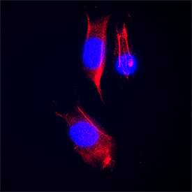
EMMPRIN/CD147 in WM‑115 Human Cell Line. EMMPRIN/CD147 was detected in immersion fixed WM‑115 human malignant melanoma cell line using Rabbit Anti-Human EMMPRIN/CD147 Monoclonal Antibody (Catalog # MAB9722) at 3 µg/mL for 3 hours at room temperature. Cells were stained using the NorthernLights™ 557-conjugated Anti-Mouse IgG Secondary Antibody (red; Catalog # NL007) and counterstained with DAPI (blue). Specific staining was localized to cytoplasm and cell membrane. View our protocol for Fluorescent ICC Staining of Cells on Coverslips.
 View Larger
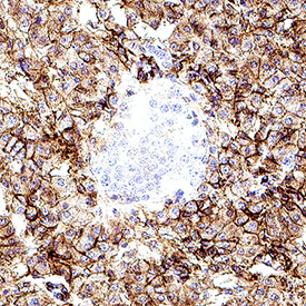
EMMPRIN/CD147 in Human Pancreas. EMMPRIN/CD147 was detected in immersion fixed paraffin-embedded sections of human pancreas using Rabbit Anti-Human EMMPRIN/CD147 Monoclonal Antibody (Catalog # MAB9722) at 3 µg/mL for 1 hour at room temperature followed by incubation with the Anti-Rabbit IgG VisUCyte™ HRP Polymer Antibody (Catalog # VC003). Before incubation with the primary antibody, tissue was subjected to heat-induced epitope retrieval using Antigen Retrieval Reagent-Basic (Catalog # CTS013). Tissue was stained using DAB (brown) and counterstained with hematoxylin (blue). Specific staining was localized to cell surface in exocrine cells. View our protocol for IHC Staining with VisUCyte HRP Polymer Detection Reagents.
Reconstitution Calculator
Preparation and Storage
- 12 months from date of receipt, -20 to -70 °C as supplied.
- 1 month, 2 to 8 °C under sterile conditions after reconstitution.
- 6 months, -20 to -70 °C under sterile conditions after reconstitution.
Background: EMMPRIN/CD147
Extracellular matrix metalloproteinase (MMP) inducer (EMMPRIN), also known as basigin and CD147, is a 44-66 kDa, variably N- and O-glycosylated, type I transmembrane protein that belongs to the immunoglobulin superfamily (1-4). Human EMMPRIN is 269 amino acids (aa) in length and contains a 24 aa signal sequence, a 183 aa extracellular domain (ECD), a 21 aa transmembrane (TM) segment and a 41 aa cytoplasmic tail. The ECD contains one C2-type and one V-type Ig-like domain. There is one 385 aa splice variant that contains an extra N-terminal IgCAM domain and is found only in the retina (5). There are additional multiple potential isoform variants for EMMMPRIN. Two that have been characterized are 205 and 176 aa in length. The 176 aa isoform utilizes an alternative start site at Met94, while the 205 aa isoform contains an 11 aa substitution for aa 1-75. Notably, the 176 aa isoform heterodimerizes with the standard EMMPRIN isoform and down-modulates its activity. This is in contrast to EMMPRIN homodimers that show full biological activity (6). EMMPRIN is expressed in areas of tissue remodeling, including endometrium, placenta, skin, and regions undergoing angiogenesis (1, 2, 7-10). It is also expressed on cells with high metabolic activity, such as lymphoblasts, macrophages and particularly tumor cells (2, 11). On such cells, EMMPRIN is often co-expressed with the amino acid transporter CD98h (12). EMMPRIN also interacts with caveolin-1 (via its C2-like domain), and this reduces the level of EMMPRIN glycosylation and subsequent EMMPRIN multimerization and activity (13). In addition, EMMMPRIN is reported to complex with both annexin II and beta 1 integrins alpha 3 and alpha 6, an interaction that contributes to tumor growth and metastasis (14-16). Finally, the soluble calcium-binding protein S100A9 has now been identified as a ligand for EMMPRIN, and may mediate many of the tumorigenic activities attributed to EMMPRIN (17). EMMPRIN's TM sequence contains a charged aa (Glu), and a Pro important for intracellular interactions with cyclophilins (CyP) (3, 18, 19). CyPA (cyclosporin A receptor) and CyP60 interactions with the TM segment promote leukocyte inflammatory chemotaxis and surface expression of EMMPRIN, respectively (18, 19). An active 22 kDa fragment can be shed from tumor cells by MT1-MMP (1). Tumor cells can also release active, full-length EMMPRIN in microvesicles (20, 21). Functionally, EMMPRIN is known to induce urokinase-type plasminogen activator (uPA), VEGF, hyaluronan and multiple MMPs (1, 2, 8-10). Human EMMPRIN (269 aa) shows 58%, 58%, 62% and 52% aa identity with mouse, rat, cow and chick EMMPRIN, respectively. It also shows 25% and 38% aa identity with the related proteins, embigin and neuroplastin (SDR-1), respectively. SARS-CoV-2 invades host cells via two receptors: angiotensin-converting enzyme 2 (ACE2) and EMMPRIN. Spike protein (SP) from virus binds to ACE2 or EMMPRIN on the host cell, mediating viral invasion and dissemination of virus among other cells (22). EMMPRIN is a second entry receptor for SARS-CoV-2 (22). It is present in multiple cellular types in lung and highly expressed in type II pneumocytes and macrophages at the edges of the fibrotic zones (22). Therefore, the blockade of EMMPRIN could also play a beneficial role in pulmonary fibrosis due to COVID-19 (22).
- Gabison, E. E. et al. (2005) Biochimie 87:361.
- Yurchenko, V. et al. (2006) Immunology 117:301.
- Kasinrerk, W. et al. (1992) J. Immunol. 149:847.
- Iacono, K.T. et al. (2007) Exp. Mol. Pathol. 83:283.
- Hanna, S. M. et al. (2003) BMC Biochem. 4:17.
- Liao, C-G. et al. (2011) Mol. Cell. Biol. 31:2591.
- Riethdorf, S. et al. (2006) Int. J. Cancer 119:1800.
- Braundmeier, A. G. et al. (2006) J. Clin. Endocrinol. Metab. 91:2358.
- Tang, Y. et al. (2006) Mol. Cancer Res. 4:371.
- Quemener, C. et al. (2007) Cancer Res. 67:9.
- Wilson, M. C. et al. (2005) J. Biol. Chem. 280:27213.
- Xu, D. and M. E. Hemler, (2005) Mol. Cell. Proteomics 4:1061.
- Tang, W. et al. (2004) Mol. Biol. Cell 15:4043.
- Zhao, P. et al. (2010) Cancer Sci. 101:387.
- Dai, J. et al. (2009) BMC Cancer 9:337.
- Li, Y. et al. (2012) J. Biol. Chem. 287:4759.
- Hibino, T. et al. (2013) Cancer Res. 73:172.
- Arora, K. et al. (2005) J. Immunol. 175:517.
- Pushkarsky, T. et al. (2005) J. Biol. Chem. 280:27866.
- Egawa, N. et al. (2006) J. Biol. Chem. 281:37576.
- Sidhu, S. S. et al. (2004) Oncogene 23:956.
- Ulrich, H. et al. (2020) Stem Cell Rev. and Rep., https://doi.org/10.1007/s12015-020-09976-7.
Product Datasheets
FAQs
No product specific FAQs exist for this product, however you may
View all Antibody FAQsReviews for Human EMMPRIN/CD147 Antibody
There are currently no reviews for this product. Be the first to review Human EMMPRIN/CD147 Antibody and earn rewards!
Have you used Human EMMPRIN/CD147 Antibody?
Submit a review and receive an Amazon gift card.
$25/€18/£15/$25CAN/¥75 Yuan/¥2500 Yen for a review with an image
$10/€7/£6/$10 CAD/¥70 Yuan/¥1110 Yen for a review without an image

