Human
EpCAM/TROP-1 Antibody Summary
Extracellular domain
Applications
Human EpCAM/TROP-1 Sandwich Immunoassay
Please Note: Optimal dilutions should be determined by each laboratory for each application. General Protocols are available in the Technical Information section on our website.
Scientific Data
 View Larger
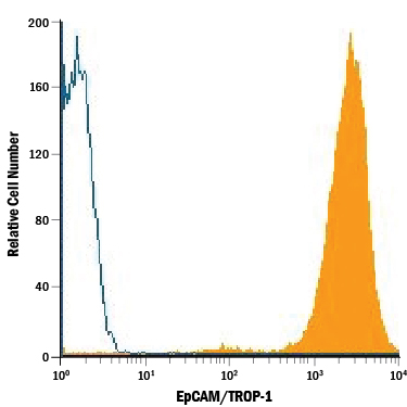
Detection of EpCAM/TROP‑1 in HT‑29 Human Cell Line by Flow Cytometry. HT-29 human colon adenocarcinoma cell line was stained with Mouse Anti-Human EpCAM/TROP-1 Monoclonal Antibody (Catalog # MAB9601, filled histogram) or isotype control antibody (Catalog # MAB0041, open histogram), followed by Phycoerythrin-conjugated Anti-Mouse IgG Secondary Antibody (Catalog # F0102B).
Reconstitution Calculator
Preparation and Storage
- 12 months from date of receipt, -20 to -70 °C as supplied.
- 1 month, 2 to 8 °C under sterile conditions after reconstitution.
- 6 months, -20 to -70 °C under sterile conditions after reconstitution.
Background: EpCAM/TROP1
Epithelial Cellular Adhesion Molecule (EpCAM), also known as KS1/4, gp40, GA733-2, 17-1A, and TROP-1, is a 40 kDa transmembrane glycoprotein composed of a 242 amino acid (aa) extracellular domain with two epidermal-growth-factor-like (EGF-like) repeats within the cysteine-rich N-terminal region, a 23 aa transmembrane domain, and a 26 aa cytoplasmic domain. Human and mouse EpCAM share 82% aa sequence identity. In human, EpCAM also shares 49% aa sequence homology with TROP-2/EGP-1. During embryonic development, EpCAM is detected in fetal lung, kidney, liver, pancreas, skin, and germ cells. In adults, human EpCAM is detected in basolateral cell membranes of all simple, pseudo-stratified, and transitional epithelia, but is not detected in normal squamous stratified epithelia, mesenchymal tissue, muscular tissue, neuro-endocrine tissue, or lymphoid tissue (1). EpCAM expression has been found to increase in actively proliferating epithelia tissues and during adult liver regeneration (1, 2). EpCAM expression is also found to increase in human malignant neoplasias, with most carcinoma expressing EpCAM including those of arising from squamousal epithelia (1). EpCAM has been shown function as a homophilic Ca2+ independent adhesion molecule (3). Homophilic adhesion via EpCAM requires the interaction of both EGF-like repeats, with the first EGF-like repeat mediating reciprocal interaction between EpCAM molecules on opposing cells, while the second repeat is involved in lateral interaction of EpCAM. Lateral interaction of EpCAM lead to the formation of dimers and tetramers (4). During homophilic adhesion the cytoplasmic tail of EpCAM interacts with the actin cytoskeleton via a direct association alpha -Actinin (5).
- Balzar, M. et al. (1999) J. Mol. Med. 77:699.
- Boer, C.J, et al. (1999) J. Pathol. 188:201.
- Litvinow, S.V. et al. (1994) J. Cell Biol. 125:437.
- Balzar, M. et al. (2001) Mol. Cell. Biol. 21:2570.
- Balzar, M. et al. (1998) Mol. Cell. Biol. 18:4388.
Product Datasheets
Citations for Human
EpCAM/TROP-1 Antibody
R&D Systems personnel manually curate a database that contains references using R&D Systems products. The data collected includes not only links to publications in PubMed, but also provides information about sample types, species, and experimental conditions.
5
Citations: Showing 1 - 5
Filter your results:
Filter by:
-
Clinical impact of different exosomes' protein expression in pancreatic ductal carcinoma patients treated with standard first line palliative chemotherapy
Authors: R Giampieri, F Piva, G Occhipinti, A Bittoni, A Righetti, S Pagliarett, A Murrone, F Bianchi, C Amantini, M Giulietti, G Ricci, G Principato, G Santoni, R Berardi, S Cascinu
PLoS ONE, 2019-05-02;14(5):e0215990.
Species: Human
Sample Types: Plasma
Applications: ELISA Detection -
Single-cell magnetic imaging using a quantum diamond microscope.
Authors: Glenn D, Lee K, Park H, Weissleder R, Yacoby A, Lukin M, Lee H, Walsworth R, Connolly C
Nat Methods, 2015-06-22;12(8):736-8.
Species: Human
Sample Types: Whole Cells
Applications: ELISA Development -
Soluble EpCAM levels in ascites correlate with positive cytology and neutralize catumaxomab activity in vitro.
Authors: Seeber A, Martowicz A, Spizzo G, Buratti T, Obrist P, Fong D, Gastl G, Untergasser G
BMC Cancer, 2015-05-07;15(0):372.
Species: Human
Sample Types: BALF
Applications: ELISA Development (Capture) -
Digital diffraction analysis enables low-cost molecular diagnostics on a smartphone.
Authors: Im H, Castro C, Shao H, Liong M, Song J, Pathania D, Fexon L, Min C, Avila-Wallace M, Zurkiya O, Rho J, Magaoay B, Tambouret R, Pivovarov M, Weissleder R, Lee H
Proc Natl Acad Sci U S A, 2015-04-13;112(18):5613-8.
Species: Human
Sample Types: Whole Cells
Applications: Immunoassay Development -
Rapid detection and profiling of cancer cells in fine-needle aspirates.
Authors: Lee H, Yoon TJ, Figueiredo JL, Swirski FK, Weissleder R
Proc. Natl. Acad. Sci. U.S.A., 2009-07-20;106(30):12459-64.
Species: Human
Sample Types: Whole Cells
Applications: Flow Cytometry
FAQs
No product specific FAQs exist for this product, however you may
View all Antibody FAQsReviews for Human
EpCAM/TROP-1 Antibody
There are currently no reviews for this product. Be the first to
review Human
EpCAM/TROP-1 Antibody and earn rewards!
Have you used Human
EpCAM/TROP-1 Antibody?
Submit a review and receive an Amazon gift card.
$25/€18/£15/$25CAN/¥75 Yuan/¥2500 Yen for a review with an image
$10/€7/£6/$10 CAD/¥70 Yuan/¥1110 Yen for a review without an image


