Human
EpCAM/TROP-1 Antibody Summary
Gln24-Lys265
Accession # CAA32870
Applications
Please Note: Optimal dilutions should be determined by each laboratory for each application. General Protocols are available in the Technical Information section on our website.
Scientific Data
 View Larger
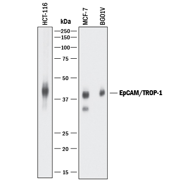
Detection of Human EpCAM/TROP‑1 by Western Blot. Western blot shows lysates of HCT-116 human colorectal carcinoma cell line, MCF-7 human breast cancer cell line, and BG01V human embryonic stem cells. PVDF membrane was probed with 0.2 µg/mL of Goat Anti-Human EpCAM/TROP-1 Antigen Affinity-purified Polyclonal Antibody (Catalog # AF960) followed by HRP-conjugated Anti-Goat IgG Secondary Antibody (HAF017). A specific band was detected for EpCAM/TROP-1 at approximately 40 kDa (as indicated). This experiment was conducted under reducing conditions and using Immunoblot Buffer Group 1.
 View Larger
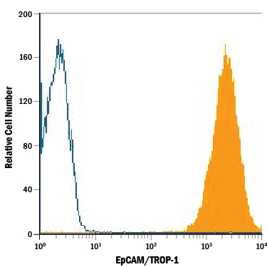
Detection of EpCAM/TROP‑1 in HT‑29 Human Cell Line by Flow Cytometry. HT-29 human colon adenocarcinoma cell line was stained with Goat Anti-Human EpCAM/TROP-1 Antigen Affinity-purified Polyclonal Antibody (Catalog # AF960, filled histogram) or isotype control antibody (AB-108-C, open histogram), followed by Phycoerythrin-conjugated Anti-Goat IgG Secondary Antibody (Catalog # F0107).
 View Larger
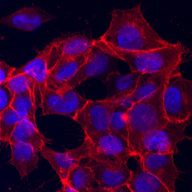
EpCAM/TROP‑1 in BG01V Human Embryonic Stem Cells. EpCAM/TROP-1 was detected in immersion fixed BG01V human embryonic stem cells using Goat Anti-Human EpCAM/TROP-1 Antigen Affinity-purified Polyclonal Antibody (Catalog # AF960) at 10 µg/mL for 3 hours at room temperature. Cells were stained using the NorthernLights™ 557-conjugated Anti-Goat IgG Secondary Antibody (red; Catalog # NL001) and counterstained with DAPI (blue). Specific staining was localized to cytoplasm and cell membrane. View our protocol for Fluorescent ICC Staining of Cells on Coverslips.
 View Larger
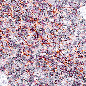
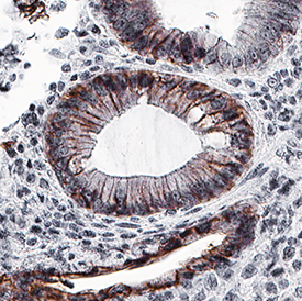
EpCAM/TROP‑1 in Human Colon. EpCAM/TROP-1 was detected in immersion fixed paraffin-embedded sections of human colon using Goat Anti-Human EpCAM/TROP-1 Antigen Affinity-purified Polyclonal Antibody (Catalog # AF960) at 0.3 µg/mL for 1 hour at room temperature followed by incubation with the Anti-Goat IgG VisUCyte™ HRP Polymer Antibody (VC004). Before incubation with the primary antibody, tissue was subjected to heat-induced epitope retrieval using Antigen Retrieval Reagent-Basic (Catalog # CTS013). Tissue was stained using DAB (brown) and counterstained with hematoxylin (blue). Specific staining was localized to cytoplasm and plasma membrane. View our protocol for IHC Staining with VisUCyte HRP Polymer Detection Reagents.
 View Larger
EpCAM/TROP‑1 in Human Colon. EpCAM/TROP-1 was detected in immersion fixed paraffin-embedded sections of human colon using Goat Anti-Human EpCAM/TROP-1 Antigen Affinity-purified Polyclonal Antibody (Catalog # AF960) at 0.3 µg/mL for 1 hour at room temperature followed by incubation with the Anti-Goat IgG VisUCyte™ HRP Polymer Antibody (VC004). Before incubation with the primary antibody, tissue was subjected to heat-induced epitope retrieval using Antigen Retrieval Reagent-Basic (CTS013). Tissue was stained using DAB (brown) and counterstained with hematoxylin (blue). Specific staining was localized to cytoplasm and plasma membrane. View our protocol for IHC Staining with VisUCyte HRP Polymer Detection Reagents.
 View Larger
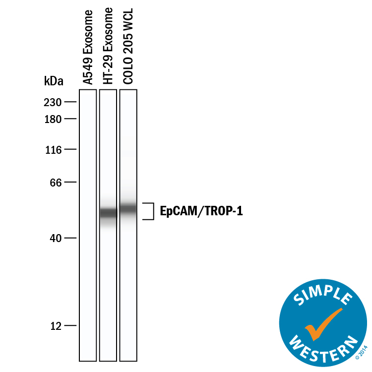
Detection of Human EpCAM/TROP‑1 by Simple WesternTM. Simple Western lane view shows lysates of A549 human lung carcinoma cell line exosomes, HT‑29 human colon adenocarcinoma cell line exosomes, and COLO 205 human colorectal adenocarcinoma cell line whole cell lysate (WCL), loaded at 0.2 mg/mL. A specific band was detected for EpCAM/TROP‑1 at approximately 52 kDa (as indicated) using 10 µg/mL of Goat Anti-Human EpCAM/TROP‑1 Antigen Affinity-purified Polyclonal Antibody (Catalog # AF960). This experiment was conducted under reducing conditions and using the 12-230 kDa separation system.
 View Larger
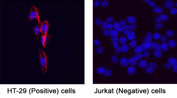
Detection of EpCAM/TROP‑1 in HT-29 Human Colon Adenocarcinoma Cell Line (Positive) and Jurkat Human Acute T Cell Leukemia Cell Line (Negative) Cells. EpCAM/TROP‑1 was detected in immersion fixed HT‑29 human colon adenocarcinoma cell line (positive) and Jurkat human acute T cell leukemia cell line (negative) cells using Goat Anti-Human EpCAM/TROP‑1 Antigen Affinity-purified Polyclonal Antibody (Catalog # AF960) at 5 µg/mL for 3 hours at room temperature. Cells were stained using the NorthernLights™ 557-conjugated Anti-Goat IgG Secondary Antibody (red; Catalog # NL001) and counterstained with DAPI (blue). Specific staining was localized to cell surface. View our protocol for Fluorescent ICC Staining of Cells on Coverslips.
Reconstitution Calculator
Preparation and Storage
- 12 months from date of receipt, -20 to -70 °C as supplied.
- 1 month, 2 to 8 °C under sterile conditions after reconstitution.
- 6 months, -20 to -70 °C under sterile conditions after reconstitution.
Background: EpCAM/TROP1
Epithelial Cellular Adhesion Molecule (EpCAM), also known as KS1/4, gp40, GA733-2, 17-1A, and TROP-1, is a 40 kDa transmembrane glycoprotein composed of a 242 amino acid (aa) extracellular domain with two epidermal-growth-factor-like (EGF-like) repeats within the cysteine-rich N-terminal region, a 23 aa transmembrane domain, and a 26 aa cytoplasmic domain. Human and mouse EpCAM share 82% aa sequence identity. In human, EpCAM also shares 49% aa sequence homology with TROP-2/EGP-1. During embryonic development, EpCAM is detected in fetal lung, kidney, liver, pancreas, skin, and germ cells. In adults, human EpCAM is detected in basolateral cell membranes of all simple, pseudo-stratified, and transitional epithelia, but is not detected in normal squamous stratified epithelia, mesenchymal tissue, muscular tissue, neuro-endocrine tissue, or lymphoid tissue (1). EpCAM expression has been found to increase in actively proliferating epithelia tissues and during adult liver regeneration (1, 2). EpCAM expression is also found to increase in human malignant neoplasias, with most carcinoma expressing EpCAM including those of arising from squamous epithelia (1). EpCAM has been shown function as a homophilic Ca2+ independent adhesion molecule (3). Homophilic adhesion via EpCAM requires the interaction of both EGF-like repeats, with the first EGF-like repeat mediating reciprocal interaction between EpCAM molecules on opposing cells, while the second repeat is involved in lateral interaction of EpCAM. Lateral interaction of EpCAM lead to the formation of dimers and tetramers (4). During homophilic adhesion the cytoplasmic tail of EpCAM interacts with the actin cytoskeleton via a direct association alpha -actinin (5).
- Balzar, M. et al. (1999) J. Mol. Med. 77:699.
- Boer, C.J. et al. (1999) J. Pathol. 188:201.
- Litvinow, S.V. et al. (1994) J. Cell Biol. 125:437.
- Balzar, M. et al. (2001) Mol. Cell. Biol. 21:2570.
- Balzar, M. et al. (1998) Mol. Cell. Biol. 18:4388.
Product Datasheets
Citations for Human
EpCAM/TROP-1 Antibody
R&D Systems personnel manually curate a database that contains references using R&D Systems products. The data collected includes not only links to publications in PubMed, but also provides information about sample types, species, and experimental conditions.
13
Citations: Showing 1 - 10
Filter your results:
Filter by:
-
Generation and expansion of transitional lung organoids from human pluripotent stem cells
Authors: IM Leko, N Schrode, J Torres, M Pezet, TA Thimraj, KG Beaumont, HW Snoeck
bioRxiv : the preprint server for biology, 2023-02-01;0(0):.
Species: Human
Sample Types: Whole Cells
Applications: ICC -
Prostate cancer extracellular vesicle digital scoring assay - a rapid noninvasive approach for quantification of disease-relevant mRNAs
Authors: JJ Wang, N Sun, YT Lee, M Kim, T Vagner, K Rohena-Riv, Z Wang, Z Chen, RY Zhang, J Lee, C Zhang, H Tang, J Widjaja, TX Zhang, D Qi, PC Teng, YJ Jan, KC Hou, C Hamann, HM Sandler, TJ Daskivich, DJ Luthringer, NA Bhowmick, R Pei, S You, DD Vizio, HR Tseng, JF Chen, Y Zhu, EM Posadas
Nano Today, 2023-01-03;48(0):.
Species: Human
Sample Types: Plasma
Applications: Capture Assay -
Targeted Inhibition of LPL/FABP4/CPT1 fatty acid metabolic axis can effectively prevent the progression of nonalcoholic steatohepatitis to liver cancer
Authors: H Yang, Q Deng, T Ni, Y Liu, L Lu, H Dai, H Wang, W Yang
International journal of biological sciences, 2021-10-11;17(15):4207-4222.
Species: Mouse
Sample Types: Whole Cells
Applications: IHC -
Immunomagnetic sequential ultrafiltration (iSUF) platform for enrichment and purification of extracellular vesicles from biofluids
Authors: J Zhang, LTH Nguyen, R Hickey, N Walters, X Wang, KJ Kwak, LJ Lee, AF Palmer, E Reátegui
Scientific Reports, 2021-04-13;11(1):8034.
Species: Human
Sample Types: Whole Cells
Applications: Flow Cytometry, ICC -
Improving hexaminolevulinate enabled cancer cell detection in liquid biopsy immunosensors
Authors: KM Chan, J Gleadle, J Li, TD Michl, K Vasilev, M MacGregor
Scientific Reports, 2021-03-31;11(1):7283.
Species: Human
Sample Types: Urine
Applications: ELISA Capture -
PAI-1-Dependent Inactivation of SMAD4-Modulated Junction and Adhesion Complex in Obese Endometrial Cancer
Authors: LL Lin, ER Kost, CL Lin, P Valente, CM Wang, MG Kolonin, AC Daquinag, X Tan, N Lucio, CN Hung, CP Wang, NB Kirma, TH Huang
Cell Rep, 2020-10-13;33(2):108253.
Species: Human
Sample Types: Whole Cells
Applications: CyTof -
Purification of HCC-specific extracellular vesicles on nanosubstrates for early HCC detection by digital scoring
Authors: N Sun, YT Lee, RY Zhang, R Kao, PC Teng, Y Yang, P Yang, JJ Wang, M Smalley, PJ Chen, M Kim, SJ Chou, L Bao, J Wang, X Zhang, D Qi, J Palomique, N Nissen, SB Han, S Sadeghi, RS Finn, S Saab, RW Busuttil, D Markovic, D Elashoff, HH Yu, H Li, AP Heaney, E Posadas, S You, JD Yang, R Pei, VG Agopian, HR Tseng, Y Zhu
Nat Commun, 2020-09-07;11(1):4489.
Species: Human
Sample Types: Plasma
Applications: ELISA Capture -
Generation of intestinal organoids derived from human pluripotent stem cells for drug testing
Authors: S Yoshida, H Miwa, T Kawachi, S Kume, K Takahashi
Sci Rep, 2020-04-06;10(1):5989.
Species: Human
Sample Types: Whole Cells
Applications: ICC -
Expansion of human primary hepatocytes in vitro through their amplification as liver progenitors in a 3D organoid system
Authors: D Garnier, R Li, F Delbos, A Fourrier, C Collet, C Guguen-Gui, C Chesné, TH Nguyen
Sci Rep, 2018-05-29;8(1):8222.
Species: Human
Sample Types: Whole Cells
Applications: ICC -
Fast and selective cell isolation from blood sample by microfiber fabric system with vacuum aspiration
Sci Technol Adv Mater, 2016-11-25;17(1):807-815.
Species: Human
Sample Types: Whole Cells
Applications: Cell Selection -
HER2 and EGFR overexpression support metastatic progression of prostate cancer to bone
Cancer Res., 2016-10-28;0(0):.
Species: Human
Sample Types: Whole Blood
Applications: CTC -
EpCAM-Regulated Transcription Exerts Influences on Nanomechanical Properties of Endometrial Cancer Cells that Promote Epithelial-to-Mesenchymal Transition
Cancer Res, 2016-08-28;0(0):.
Species: Human
Sample Types: Whole Cells
Applications: Adhesion Assay -
Stromal gene expression defines poor-prognosis subtypes in colorectal cancer.
Authors: Calon A, Lonardo E, Berenguer-Llergo A, Espinet E, Hernando-Momblona X, Iglesias M, Sevillano M, Palomo-Ponce S, Tauriello D, Byrom D, Cortina C, Morral C, Barcelo C, Tosi S, Riera A, Attolini C, Rossell D, Sancho E, Batlle E
Nat Genet, 2015-02-23;47(4):320-9.
Species: Human
Sample Types: Whole Tissue
Applications: IHC
FAQs
No product specific FAQs exist for this product, however you may
View all Antibody FAQsReviews for Human
EpCAM/TROP-1 Antibody
There are currently no reviews for this product. Be the first to
review Human
EpCAM/TROP-1 Antibody and earn rewards!
Have you used Human
EpCAM/TROP-1 Antibody?
Submit a review and receive an Amazon gift card.
$25/€18/£15/$25CAN/¥75 Yuan/¥2500 Yen for a review with an image
$10/€7/£6/$10 CAD/¥70 Yuan/¥1110 Yen for a review without an image


