Human EphA2 Antibody Summary
Gln25-Asn534
Accession # P29317
Applications
Please Note: Optimal dilutions should be determined by each laboratory for each application. General Protocols are available in the Technical Information section on our website.
Scientific Data
 View Larger
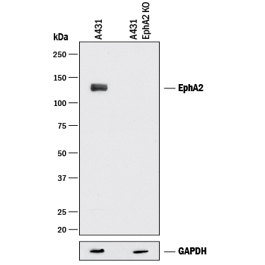
Detection of Human EphA2 by Western Blot. Western blot shows lysates of A431 human epithelial carcinoma parental cell line and EphA2 knock out (KO) A431 cell line. PVDF membrane was probed with 1 µg/mL of Goat Anti-Human EphA2 Antigen Affinity-purified Polyclonal Antibody (Catalog # AF3035) followed by HRP-conjugated Anti-Goat IgG Secondary Antibody (HAF017). A specific band was detected for EphA2 at approximately 110 kDa (as indicated), but not detectable in the knockout A431 cell line. GAPDH (Catalog # MAB5718) is shown as a loading control. This experiment was conducted under reducing conditions and using Immunoblot Buffer Group 1.
 View Larger
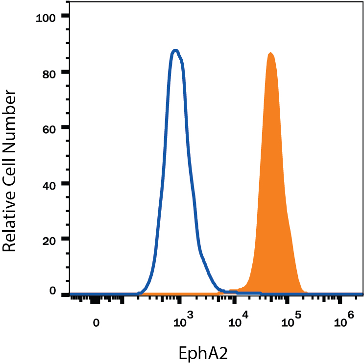
Detection of EphA2 in A431 Human Cell Line by Flow Cytometry. A431 human epithelial carcinoma cell line was stained with Goat Anti-Human EphA2 Antigen Affinity-purified Polyclonal Antibody (Catalog # AF3035, filled histogram) or isotype control antibody (Catalog # AB-108-C, open histogram), followed by Phycoerythrin-conjugated Anti-Goat IgG Secondary Antibody (Catalog # F0107). View our protocol for Staining Membrane-associated Proteins.
 View Larger
EphA2 in Human Ovarian Cancer Tissue. EphA2 was detected in immersion fixed paraffin-embedded sections of human ovarian cancer tissue using Goat Anti-Human EphA2 Antigen Affinity-purified Polyclonal Antibody (Catalog # AF3035) at 5 µg/mL overnight at 4 °C. Tissue was stained using the Anti-Goat HRP-DAB Cell & Tissue Staining Kit (brown; Catalog # CTS008) and counterstained with hematoxylin (blue). Specific labeling was localized to the plasma membrane of cancer cells. View our protocol for Chromogenic IHC Staining of Paraffin-embedded Tissue Sections.
Reconstitution Calculator
Preparation and Storage
- 12 months from date of receipt, -20 to -70 °C as supplied.
- 1 month, 2 to 8 °C under sterile conditions after reconstitution.
- 6 months, -20 to -70 °C under sterile conditions after reconstitution.
Background: EphA2
EphA2, also known as Eck, Myk2, and Sek2, is a member of the Eph receptor tyrosine kinase family which binds Ephrins A1, 2, 3, 4, and 5 (1-4). A and B class Eph proteins have a common structural organization. The human EphA2 cDNA encodes a 976 amino acid (aa) precursor including a 24 aa signal sequence, a 510 aa extracellular domain (ECD), a 24 aa transmembrane segment, and a 418 aa cytoplasmic domain. The ECD contains an N-terminal globular domain, a cysteine-rich domain, and two fibronectin type III domains (5). The cytoplasmic domain contains a juxtamembrane motif with two tyrosine residues, which are the major autophosphorylation sites, a kinase domain, and a sterile alpha motif (SAM) (5). The ECD of human EphA2 shares 90-94% aa sequence identity with mouse, bovine, and canine EphA2, and approximately 45% aa sequence identity with human EphA1, 3, 4, 5, 7, and 8. EphA2 becomes autophosphorylated following ligand binding (6, 7) and then interacts with SH2 domain-containing PI3-kinase to activate MAPK pathways (8, 9). Reverse signaling is also propagated through the Ephrin ligand. Transcription of EphA2 is dependent on the expression of E-Cadherin (10), and can be induced by p53 family transcription factors (11). EphA2 is upregulated in breast, prostate, and colon cancer vascular endothelium. Its ligand, EphrinA1, is expressed by the local tumor cells (12, 13). In some cases, EphA2 and EphrinA1 are expressed on the same blood vessels (14). EphA2 signaling cooperates with VEGF receptor signaling in promoting endothelial cell migration (13). The gene encoding human EphA2 maps to a region on chromosome 1 which is frequently deleted in neuroectodermal tumors (15).
- Poliakov, A. et al. (2004) Dev. Cell 7:465.
- Surawska, H. et al. (2004) Cytokine Growth Factor Rev. 15:419.
- Pasquale, E.B. (2005) Nat. Rev. Mol. Cell Biol. 6:462.
- Davy, A. and P. Soriano (2005) Dev. Dyn. 232:1.
- Bohme, B et al. (1993) Oncogene 8:2857.
- Pandey, A. et al. (1995) Science 268:567.
- Bartley, T.D. et al. (1994) Nature 368:558.
- Pandey, A. et al. (1994) J. Biol. Chem. 269:30154.
- Miao, H. et al. (2001) Nat. Cell Biol. 3:527.
- Orsulic, S. and R. Kemler (2000) J. Cell Sci. 113:1793.
- Dohn, M. et al. (2001) Oncogene 20:6503.
- Zelinski, D.P. et al. (2001) Cancer Res. 61:2301.
- Brantley, D.M. et al. (2002) Oncogene 21:7011.
- Ogawa, K. et al. (2000) Oncogene 19:6043.
- Sulman, E.P. et al. (1997) Genomics 40:371.
Product Datasheets
Citations for Human EphA2 Antibody
R&D Systems personnel manually curate a database that contains references using R&D Systems products. The data collected includes not only links to publications in PubMed, but also provides information about sample types, species, and experimental conditions.
11
Citations: Showing 1 - 10
Filter your results:
Filter by:
-
Identification of potent pan-ephrin receptor kinase inhibitors using DNA-encoded chemistry technology
Authors: Madasu, C;Liao, Z;Parks, SE;Sharma, KL;Bohren, KM;Ye, Q;Li, F;Palaniappan, M;Tan, Z;Yuan, F;Creighton, CJ;Tang, S;Masand, RP;Guan, X;Young, DW;Monsivais, D;Matzuk, MM;
Proceedings of the National Academy of Sciences of the United States of America
Species: Human
Sample Types: Whole Tissue
Applications: Immunohistochemistry -
Aggressive and recurrent ovarian cancers upregulate ephrinA5, a non-canonical effector of EphA2 signaling duality
Authors: J Jukonen, L Moyano-Gal, K Höpfner, EA Pietilä, L Lehtinen, K Huhtinen, E Gucciardo, J Hynninen, S Hietanen, S Grénman, PM Ojala, O Carpén, K Lehti
Scientific Reports, 2021-04-23;11(1):8856.
Species: Human
Sample Types: Cell Lysates, Whole Cells, Whole Tissue
Applications: ICC, IHC, Western Blot -
MyD88/CD40 signaling retains CAR T cells in a less differentiated state
Authors: B Prinzing, P Schreiner, M Bell, Y Fan, G Krenciute, S Gottschalk
JCI Insight, 2020-11-05;5(21):.
Species: Human
Sample Types: Cell Lysates
Applications: Western Blot -
Adaptive RSK-EphA2-GPRC5A signaling switch triggers chemotherapy resistance in ovarian cancer
Authors: L Moyano-Gal, EA Pietilä, SP Turunen, S Corvigno, E Hjerpe, D Bulanova, U Joneborg, T Alkasalias, Y Miki, M Yashiro, A Chernenko, J Jukonen, M Singh, H Dahlstrand, JW Carlson, K Lehti
EMBO Mol Med, 2020-03-02;0(0):e11177.
Species: Human
Sample Types: Cell Culture Lysates
Applications: ICC -
Stemness underpinning all steps of human colorectal cancer defines the core of effective therapeutic strategies
Authors: A Visioli, F Giani, N Trivieri, R Pracella, E Miccinilli, MG Cariglia, O Palumbo, A Arleo, F Dezi, M Copetti, L Cajola, S Restelli, V Papa, A Sciuto, TP Latiano, M Carella, D Amadori, G Gallerani, R Ricci, S Alfieri, G Pesole, AL Vescovi, E Binda
EBioMedicine, 2019-05-02;0(0):.
Species: Human
Sample Types: Whole Cells
Applications: ICC -
Brucine Suppresses Vasculogenic Mimicry in Human Triple-Negative Breast Cancer Cell Line MDA-MB-231
Authors: MR Xu, PF Wei, MZ Suo, Y Hu, W Ding, L Su, YD Zhu, WJ Song, GH Tang, M Zhang, P Li
Biomed Res Int, 2019-01-06;2019(0):6543230.
Species: Human
Sample Types: Cell Lysates
Applications: Western Blot -
Engineered nanointerfaces for microfluidic isolation and molecular profiling of tumor-specific extracellular vesicles
Authors: E Reátegui, KE van der Vo, CP Lai, M Zeinali, NA Atai, B Aldikacti, FP Floyd, A H Khankhel, V Thapar, FH Hochberg, LV Sequist, BV Nahed, B S Carter, M Toner, L Balaj, D T Ting, XO Breakefiel, SL Stott
Nat Commun, 2018-01-12;9(1):175.
Species: Human
Sample Types: Whole Cells
Applications: Functional Assay -
Protein kinase A can block EphA2 receptor-mediated cell repulsion by increasing EphA2 S897 phosphorylation.
Authors: Barquilla A, Lamberto I, Heynen-Genel S, Brill L, Pasquale E
Mol Biol Cell, 2016-07-06;27(17):2757-70.
Species: Human
Sample Types: Cell Lysates
Applications: Western Blot -
EphA2 cleavage by MT1-MMP triggers single cancer cell invasion via homotypic cell repulsion.
Authors: Sugiyama N, Gucciardo E, Tatti O, Varjosalo M, Hyytiainen M, Gstaiger M, Lehti K
J Cell Biol, 2013-04-29;201(3):467-84.
Species: Human, Primate
Sample Types: Cell Lysates
Applications: Western Blot -
Alteration of the EphA2/Ephrin-A signaling axis in psoriatic epidermis.
Authors: Gordon K, Kochkodan J, Blatt H, Lin S, Kaplan N, Johnston A, Swindell W, Hoover P, Schlosser B, Elder J, Gudjonsson J, Getsios S
J Invest Dermatol, 2012-11-29;133(3):712-22.
Species: Human
Sample Types: Whole Tissue
Applications: IHC-Fr -
A novel extracellular Hsp90 mediated co-receptor function for LRP1 regulates EphA2 dependent glioblastoma cell invasion.
Authors: Gopal U, Bohonowych JE, Lema-Tome C
PLoS ONE, 2011-03-08;6(3):e17649.
Species: Human
Sample Types: Whole Cells, Whole Tissue
Applications: Flow Cytometry, IHC-P
FAQs
No product specific FAQs exist for this product, however you may
View all Antibody FAQsReviews for Human EphA2 Antibody
There are currently no reviews for this product. Be the first to review Human EphA2 Antibody and earn rewards!
Have you used Human EphA2 Antibody?
Submit a review and receive an Amazon gift card.
$25/€18/£15/$25CAN/¥75 Yuan/¥2500 Yen for a review with an image
$10/€7/£6/$10 CAD/¥70 Yuan/¥1110 Yen for a review without an image

