Human Fas Ligand/TNFSF6 Antibody Summary
Pro134-Leu281
Accession # P48023
Applications
Please Note: Optimal dilutions should be determined by each laboratory for each application. General Protocols are available in the Technical Information section on our website.
Scientific Data
 View Larger
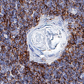
Fas Ligand/TNFSF6 in Human Thymus. Fas Ligand/TNFSF6 was detected in immersion fixed paraffin-embedded sections of human thymus (Hassall's corpuscle) using Mouse Anti-Human Fas Ligand/TNFSF6 Monoclonal Antibody (Catalog # MAB0951) at 5 µg/mL for 1 hour at room temperature followed by incubation with the Anti-Mouse IgG VisUCyte™ HRP Polymer Antibody (Catalog # VC001). Before incubation with the primary antibody, tissue was subjected to heat-induced epitope retrieval using Antigen Retrieval Reagent-Basic (Catalog # CTS013). Tissue was stained using DAB (brown) and counterstained with hematoxylin (blue). Specific staining was localized to cytoplasm. View our protocol for IHC Staining with VisUCyte HRP Polymer Detection Reagents.
Reconstitution Calculator
Preparation and Storage
- 12 months from date of receipt, -20 to -70 °C as supplied.
- 1 month, 2 to 8 °C under sterile conditions after reconstitution.
- 6 months, -20 to -70 °C under sterile conditions after reconstitution.
Background: Fas Ligand/TNFSF6
Fas Ligand (FasL), also known as CD178, CD95L, or TNFSF6, is a 40 kDa type II transmembrane member of the TNF superfamily of proteins. Its ability to induce apoptosis in target cells plays an important role in the development, homeostasis, and function of the immune system (1). Mature human Fas Ligand consists of a 179 amino acid (aa) extracellular domain (ECD), a 22 aa transmembrane segment, and a 80 aa cytoplasmic domain (2). Within the ECD, human Fas Ligand shares 81% and 78% aa sequence identity with mouse and rat Fas Ligand, respectively. Both mouse and human Fas Ligand are active on mouse and human cells (2, 3). Fas Ligand is expressed on the cell surface as a nondisulfide-linked homotrimer on activated CD4+ Th1 cells, CD8+ cytotoxic T cells, and NK cells (1). Fas Ligand binding to Fas/CD95 on an adjacent cell triggers apoptosis in the Fas‑expressing cell (2, 4). Fas Ligand also binds DcR3 which is a soluble decoy receptor that interferes with Fas Ligand-induced apoptosis (5). Fas Ligand can be released from the cell surface by metalloproteinases as a 26 kDa soluble molecule which remains trimeric (6, 7). Shed Fas Ligand retains the ability to bind Fas, although its ability to trigger apoptosis is dramatically reduced (6, 7). In the absence of TGF‑ beta, however, Fas Ligand/Fas interactions instead promote neutrophil-mediated inflammatory responses (3, 8). Fas Ligand itself transmits reverse signals that costimulate the proliferation of freshly antigen-stimulated T cells (9). Fas Ligand-induced apoptosis plays a central role in the development of immune tolerance and the maintance of immune privileged sites (10). This function is exploited by tumor cells which evade immune surveillance by upregulating Fas Ligand to kill tumor infiltrating lymphocytes (8, 11). In gld mice, a Fas Ligand point mutation is the cause of severe lymphoproliferation and systemic autoimmunity (12, 13).
- Lettau, M. et al. (2008) Curr. Med. Chem. 15:1684.
- Takahashi, T. et al. (1994) Int. Immunol. 6:1567.
- Seino, K-I. et al. (1998) J. Immunol. 161:4484.
- Suda, T. et al. (1993) Cell 75:1169.
- Pitti, R.M. et al. (1998) Nature 396:699.
- Schneider, P. et al. (1998) J. Exp. Med. 187:1205.
- Tanaka, M. et al. (1998) Nature Med. 4:31.
- Chen, J-J. et al. (1998) Science 282:1714.
- Suzuki, I. and P.J. Fink (2000) Proc. Natl. Acad. Sci. 97:1707.
- Ferguson, T.A. and T.S. Griffith (2006) Immunol. Rev. 213:228.
- Ryan, A.E. et al. (2005) Cancer Res. 65:9817.
- Takahashi, T. et al. (1994) Cell 76:969.
- Lynch, D.H. et al. (1994) Immunity 1:131.
Product Datasheets
FAQs
No product specific FAQs exist for this product, however you may
View all Antibody FAQsReviews for Human Fas Ligand/TNFSF6 Antibody
Average Rating: 5 (Based on 1 Review)
Have you used Human Fas Ligand/TNFSF6 Antibody?
Submit a review and receive an Amazon gift card.
$25/€18/£15/$25CAN/¥75 Yuan/¥2500 Yen for a review with an image
$10/€7/£6/$10 CAD/¥70 Yuan/¥1110 Yen for a review without an image
Filter by:


