Human FoxP1 Antibody Summary
Lys548-Glu677
Accession # Q9H334
Applications
Please Note: Optimal dilutions should be determined by each laboratory for each application. General Protocols are available in the Technical Information section on our website.
Scientific Data
 View Larger
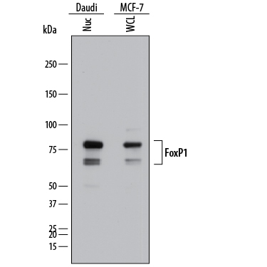
Detection of Human FoxP1 by Western Blot. Western blot shows lysates of Daudi human Burkitt's lymphoma cell line and MCF-7 human breast cancer cell line. Gels were loaded with 25 µg of whole cell lysate (WCL) and 25 µg of nuclear extracts (Nuc). PVDF membrane was probed with 1 µg/mL of Mouse Anti-Human FoxP1 Monoclonal Antibody (Catalog # MAB45341) followed by HRP-conjugated Anti-Mouse IgG Secondary Antibody (Catalog # HAF018). Specific bands were detected for FoxP1 at approximately 65 and 80 kDa (as indicated). This experiment was conducted under reducing conditions and using Immunoblot Buffer Group 1.
 View Larger
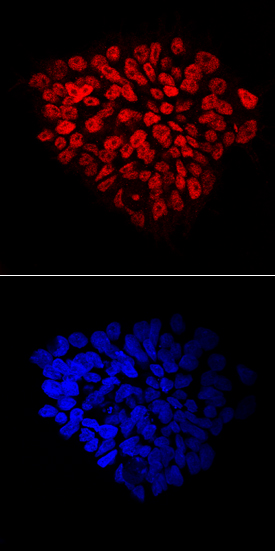
FoxP1 in BG01V Human Embryonic Stem Cells. FoxP1 was detected in immersion fixed BG01V human embryonic stem cells using Mouse Anti-Human FoxP1 Monoclonal Antibody (Catalog # MAB45341) at 10 µg/mL for 3 hours at room temperature. Cells were stained using the NorthernLights™ 557-conjugated Anti-Mouse IgG Secondary Antibody (red, upper panel; Catalog # NL007) and counterstained with DAPI (blue, lower panel). Specific staining was localized to nuclei. View our protocol for Fluorescent ICC Staining of Stem Cells on Coverslips.
 View Larger
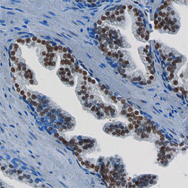
FoxP1 in Human Prostate. FoxP1 was detected in immersion fixed paraffin-embedded sections of human prostate using Mouse Anti-Human FoxP1 Monoclonal Antibody (Catalog # MAB45341) at 15 µg/mL overnight at 4 °C. Before incubation with the primary antibody, tissue was subjected to heat-induced epitope retrieval using Antigen Retrieval Reagent-Basic (Catalog # CTS013). Tissue was stained using the Anti-Mouse HRP-DAB Cell & Tissue Staining Kit (brown; Catalog # CTS002) and counter-stained with hematoxylin (blue). Specific staining was localized to the nuclei of epithelial cells. View our protocol for Chromogenic IHC Staining of Paraffin-embedded Tissue Sections.
 View Larger
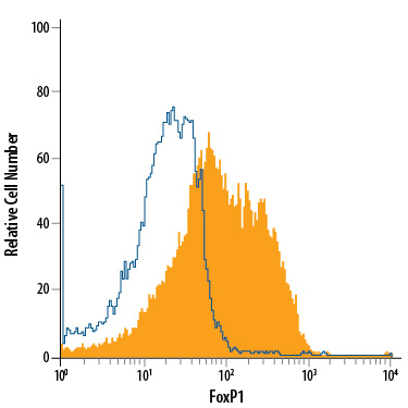
Detection of FoxP1 in MCF‑7 Human Cell Line by Flow Cytometry. MCF-7 human breast cancer cell line was stained with Mouse Anti-Human FoxP1 Monoclonal Antibody (Catalog # MAB45341, filled histogram) or isotype control antibody (Catalog # MAB002, open histogram), followed by Allophycocyanin-conjugated Anti-Mouse IgG Secondary Antibody (Catalog # F0101B). To facilitate intracellular staining, cells were fixed with 4% paraformaldehyde and permeabilized with methanol.
 View Larger
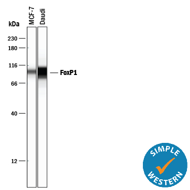
Detection of Human FoxP1 by Simple WesternTM. Simple Western lane view shows lysates of MCF‑7 human breast cancer cell line and Daudi human Burkitt's lymphoma cell line, loaded at 0.5 mg/mL. A specific band was detected for FoxP1 at approximately 99 kDa (as indicated) using 10 µg/mL of Mouse Anti-Human FoxP1 Monoclonal Antibody (Catalog # MAB45341). This experiment was conducted under reducing conditions and using the 12-230 kDa separation system.
Reconstitution Calculator
Preparation and Storage
- 12 months from date of receipt, -20 to -70 °C as supplied.
- 1 month, 2 to 8 °C under sterile conditions after reconstitution.
- 6 months, -20 to -70 °C under sterile conditions after reconstitution.
Background: FoxP1
Forkhead Box P1 (FOXP1) is a member of the FOX family of transcription factors. FoxP1 has been implicated in cardiac, lung, and lymphocyte development. FoxP1 knock out mice die at embryonic day 14.5 due to heart valve and outflow tract abnormalities. FoxP1 contains both a DNA binding domain as well as protein-protein interaction domains. FoxP1 can homo or heterodimerize with FoxP2 and FoxP4, with dimerization necessary for DNA binding. FoxP1 shows both oncogenic and tumor suppressive characteristics. Overexpression in lymphomas leads to poor prognosis, but loss of FoxP1 in breast cancer also implicates a poor prognosis. Human isoforms of 489 to 677 amino acids contain alternate sequences within the first 60 amino acids and/or deletion of amino acids.
Product Datasheets
Citations for Human FoxP1 Antibody
R&D Systems personnel manually curate a database that contains references using R&D Systems products. The data collected includes not only links to publications in PubMed, but also provides information about sample types, species, and experimental conditions.
2
Citations: Showing 1 - 2
Filter your results:
Filter by:
-
Cell cycle inhibitors protect motor neurons in an organoid model of Spinal Muscular Atrophy
Authors: JH Hor, ES Soh, LY Tan, VJW Lim, MM Santosa, Winanto, BX Ho, Y Fan, BS Soh, SY Ng
Cell Death Dis, 2018-10-27;9(11):1100.
Species: Human
Sample Types: Organoids
Applications: IHC -
Regulated expression of the TPbeta isoform of the human T prostanoid receptor by the tumour suppressors FOXP1 and NKX3.1: Implications for the role of thromboxane in prostate cancer.
Authors: O'Sullivan A, Eivers S, Mulvaney E, Kinsella B
Biochim Biophys Acta Mol Basis Dis, 2017-09-08;1863(12):3153-3169.
Species: Human
Sample Types: Cell Lysates
Applications: Western Blot
FAQs
No product specific FAQs exist for this product, however you may
View all Antibody FAQsReviews for Human FoxP1 Antibody
There are currently no reviews for this product. Be the first to review Human FoxP1 Antibody and earn rewards!
Have you used Human FoxP1 Antibody?
Submit a review and receive an Amazon gift card.
$25/€18/£15/$25CAN/¥75 Yuan/¥2500 Yen for a review with an image
$10/€7/£6/$10 CAD/¥70 Yuan/¥1110 Yen for a review without an image

