Human Galectin-10 Antibody Summary
Ser2-Arg142
Accession # Q05315
Applications
Please Note: Optimal dilutions should be determined by each laboratory for each application. General Protocols are available in the Technical Information section on our website.
Scientific Data
 View Larger
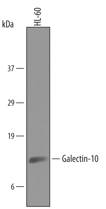
Detection of Human Galectin‑10 by Western Blot. Western blot shows lysates of HL-60 human acute promyelocytic leukemia cell line. PVDF membrane was probed with 1 µg/mL of Goat Anti-Human Galectin-10 Antigen Affinity-purified Polyclonal Antibody (Catalog # AF5447) followed by HRP-conjugated Anti-Goat IgG Secondary Antibody (Catalog # HAF019). A specific band was detected for Galectin-10 at approximately 16 kDa (as indicated). This experiment was conducted under reducing conditions and using Immunoblot Buffer Group 8.
 View Larger
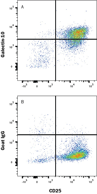
Detection of Galectin‑10 in Human Tregs by Flow Cytometry. Human Tregs were expanded from PBMC using Cloudz Human Treg Expansion Kit (CLD006) then stained with (A) Goat Anti-Human Galectin-10 Antigen Affinity-purified Polyclonal Antibody (Catalog # AF5447) or (B) Goat IgG Control Antibody (AB-108-C) followed by Phycoerythrin-conjugated Anti-Goat IgG Secondary Antibody (F0107) and Mouse Anti-Human CD25/IL-2 Ra Allophycocyanin-conjugated Monoclonal Antibody (FAB1020A). To facilitate intracellular staining, cells were fixed and permeabilized with FlowX FoxP3/Transcription Factor Fixation & Perm Buffer Kit (FC012). Staining was performed using our Staining Intracellular Molecules protocol.
 View Larger
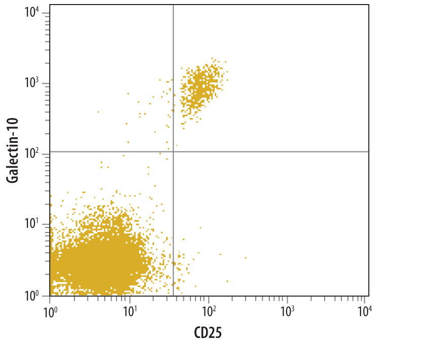
Detection of Galectin‑10 in Human PBMC lymphocytes by Flow Cytometry. Human PBMC lymphocytes were stained with Goat Anti-Human Galectin-10 Antigen Affinity-purified Polyclonal Antibody (Catalog # AF5447) followed by CFS-conjugated Anti-Goat IgG Secondary Antibody (Catalog # F0109) and Mouse Anti-Human CD25/IL-2 Ra Allophycocyanin-conjugated Monoclonal Antibody (Catalog # FAB1020A).Quadrant markers were set based on control antibody staining (Catalog # AB-108-C).
Reconstitution Calculator
Preparation and Storage
- 12 months from date of receipt, -20 to -70 °C as supplied.
- 1 month, 2 to 8 °C under sterile conditions after reconstitution.
- 6 months, -20 to -70 °C under sterile conditions after reconstitution.
Background: Galectin-10
Galectin-10 (also eosinophil lysophospholipase and Charcot-Leyden Crystal protein) is a 16 kDa member of the lectin family of proteins. It is expressed intracellularly by eosinophils, basophils and CD25+ Treg cells. Although originally believed to possess lysophospholipase activity, this has been shown to be incorrect. It is known to bind lysophospholipase and its inhibitors and to bind mannose in a very unusual manner. Human Galectin-10 is 142 amino acids (aa) in length. There is one Galectin domain (aa 6‑138) that contains two dimerization motifs (aa 6‑10 and 131‑135). Two molecular weight isoforms of 15 and 14 kDa have been described that are not yet characterized. There is no known structural rodent counterpart to human Galectin-10.
Product Datasheets
Citation for Human Galectin-10 Antibody
R&D Systems personnel manually curate a database that contains references using R&D Systems products. The data collected includes not only links to publications in PubMed, but also provides information about sample types, species, and experimental conditions.
1 Citation: Showing 1 - 1
-
Galectin-10, a potential biomarker of eosinophilic airway inflammation.
Authors: Chua, Justin C, Douglass, Jo A, Gillman, Andrew
PLoS ONE, 2012-08-06;7(8):e42549.
Species: Human
Sample Types: Sputum
Applications: Western Blot
FAQs
No product specific FAQs exist for this product, however you may
View all Antibody FAQsReviews for Human Galectin-10 Antibody
There are currently no reviews for this product. Be the first to review Human Galectin-10 Antibody and earn rewards!
Have you used Human Galectin-10 Antibody?
Submit a review and receive an Amazon gift card.
$25/€18/£15/$25CAN/¥75 Yuan/¥2500 Yen for a review with an image
$10/€7/£6/$10 CAD/¥70 Yuan/¥1110 Yen for a review without an image

