Human GluR2 Antibody Summary
Accession # P42262
Applications
Please Note: Optimal dilutions should be determined by each laboratory for each application. General Protocols are available in the Technical Information section on our website.
Scientific Data
 View Larger
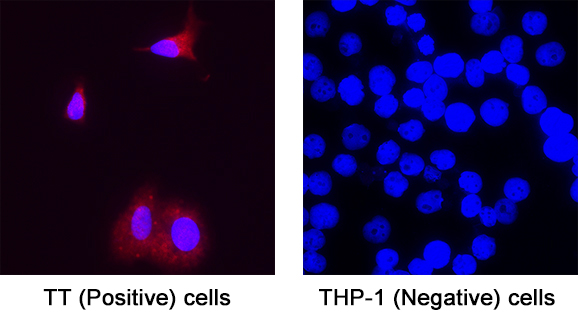
Detection of GluR2 in TT (Positive) and THP‑1 (Negative) cells. GluR2 was detected in immersion fixed TT Human Medullary Thyroid Cancer Cells (Positive) and absent in THP‑1 Human Acute Monocytic Leukemia Cells (Negative) using Mouse Anti-Human GluR2 Monoclonal Antibody (Catalog # MAB11333) at 8 µg/mL for 3 hours at room temperature. Cells were stained using the NorthernLights™ 557-conjugated Anti-Mouse IgG Secondary Antibody (red; Catalog # NL007) and counterstained with DAPI (blue). Specific staining was localized to cytoplasm. View our protocol for Fluorescent ICC Staining of Cells on Coverslips.
 View Larger
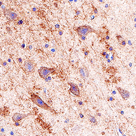
Detection of GluR2 in Human Hippocampus. GluR2 was detected in immersion fixed paraffin-embedded sections of Human Hippocampus using Mouse Anti-Human GluR2 Monoclonal Antibody (Catalog # MAB11333) at 5 µg/mL for 1 hour at room temperature followed by incubation with the Anti-Mouse IgG VisUCyte™ HRP Polymer Antibody (Catalog # VC001). Before incubation with the primary antibody, tissue was subjected to heat-induced epitope retrieval using VisUCyte Antigen Retrieval Reagent-Basic (Catalog # VCTS021). Tissue was stained using DAB (brown) and counterstained with hematoxylin (blue). Specific staining was localized to plasma membrane and cytoplasm in neurons. View our protocol for IHC Staining with VisUCyte HRP Polymer Detection Reagents.
Reconstitution Calculator
Preparation and Storage
- 12 months from date of receipt, -20 to -70 °C as supplied.
- 1 month, 2 to 8 °C under sterile conditions after reconstitution.
- 6 months, -20 to -70 °C under sterile conditions after reconstitution.
Background: GluR2
GluR2 is a receptor for glutamate. Glutamate receptors are the predominant excitatory neurotransmitter receptors in the mammalian brain. The subunit encoded by this gene (GRIA2) is subject to RNA editing which renders the receptor it becomes part of impermeable to calcium ions. This editing occurs at the Q/R site at a frequency of 100% of GluR2 transcripts in the brain. The main function of this editing is regulation of electrophysiology of the receptor channel. Defective editing of this channel has been linked to several conditions such as ALS, Epilepsy and some human brain tumors. GRIA2 is a diagnostic immunochemical marker for solitary fibrous tumor which distinguishes it from most mimics.
- Entrez Gene: GRIA glutamate receptor, ionotropic, AMPA 2.
- Seeburg PH, Single F, Kuner T, Higuchi M, Sprengel R. "Genetic Manipulation of Key Determinants of Ion Flow in Glutamate Receptor Channels in the Mouse". Brain Res. 2001 Jul; 907(1-2):233.
- Cleveland DW, Rothstein JD. "From Charcot to Lou Gehrig: Deciphering Selective Motor Neuron Death in ALS". Nat. Rev. Neurosci. 2001 Nov; 2(11):806.
- Maas S, Patt S, Schrey M, Rich A. "Underediting of Glutamate Receptor GluR-B mRNA in Malignant Gliomas". Proc. Natl. Acad. Sci. U.S.A. 2001 Dec; 98(25):14687.
- Vivero M, Doyle L.A, Fletcher C.D., Mertens F, Hornick J.L. "GRIA2 is a Novel Diagnostic Marker for Solitary Fibrous Tumour Identified through Gene Expression Profiling". Histopathology. 2014. 65(1):71.
Product Datasheets
FAQs
No product specific FAQs exist for this product, however you may
View all Antibody FAQsReviews for Human GluR2 Antibody
There are currently no reviews for this product. Be the first to review Human GluR2 Antibody and earn rewards!
Have you used Human GluR2 Antibody?
Submit a review and receive an Amazon gift card.
$25/€18/£15/$25CAN/¥75 Yuan/¥1250 Yen for a review with an image
$10/€7/£6/$10 CAD/¥70 Yuan/¥1110 Yen for a review without an image

