Human Glut1 Antibody Summary
Met1-Val492
Accession # AAA52571
Applications
Please Note: Optimal dilutions should be determined by each laboratory for each application. General Protocols are available in the Technical Information section on our website.
Scientific Data
 View Larger
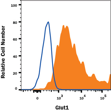
Detection of Glut1 in HepG2 Human Cell Line by Flow Cytometry. HepG2 human hepatocellular carcinoma cell line was stained with Mouse Anti-Human Glut1 Monoclonal Antibody (Catalog # MAB1418, filled histogram) or isotype control antibody (Catalog # MAB0041, open histogram), followed by Allophycocyanin-conjugated Anti-Mouse IgG Secondary Antibody (Catalog # F0101B).
 View Larger
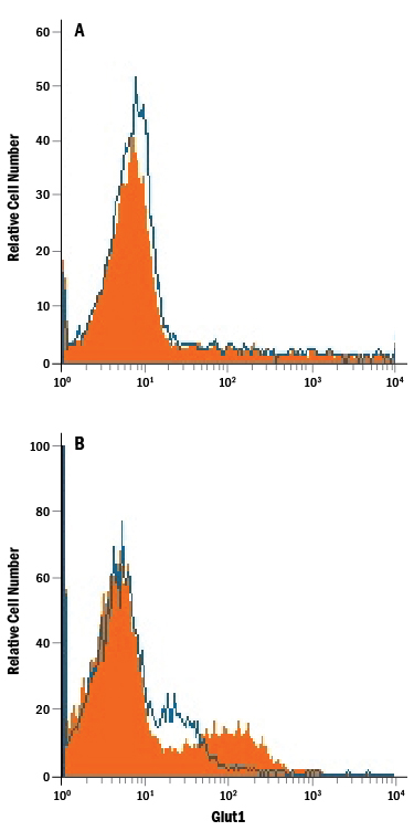
Detection of Glut1 in Jurkat Human Cell Line by Flow Cytometry. Jurkat human acute T cell leukemia cell line either (A) untreated or (B) cultured in nutrient-depleted media was stained with Mouse Anti-Human Glut1 Monoclonal Antibody (Catalog # MAB1418, filled histogram) or isotype control antibody (Catalog # MAB0041, open histogram), followed by Phycoerythrin-conjugated Anti-Mouse IgG Secondary Antibody (Catalog # F0102B).
 View Larger
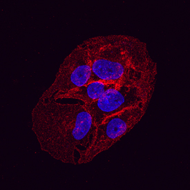
Glut1 in HepG2 Human Hepatocellular Carcinoma Cell Line. Glut1 was detected in immersion fixed HepG2 human hepatocellular carcinoma cell line using Mouse Anti-Human Glut1 Monoclonal Antibody (Catalog # MAB1418) at 10 µg/mL for 3 hours at room temperature. Cells were stained using the NorthernLights™ 557-conjugated Anti-Mouse IgG Secondary Antibody (red; Catalog # NL007) and counterstained with DAPI(blue). Specific staining was localized to the plasma membrane. View our protocol for Fluorescent ICC Staining of Cells on Coverslips.
Reconstitution Calculator
Preparation and Storage
- 12 months from date of receipt, -20 to -70 °C as supplied.
- 1 month, 2 to 8 °C under sterile conditions after reconstitution.
- 6 months, -20 to -70 °C under sterile conditions after reconstitution.
Background: Glut1
Glut1 belongs to the facilitative glucose transport protein family that comprises 13 members. It is an integral membrane protein with 12 transmembrane domains and is expressed at variable levels in many tissues including brain endothelial cells, CD8+ T cells, and erythrocytes (1‑4). Glut1 is a major glucose transporter that mediates glucose transport across the mammalian blood‑brain barrier.
- Mueckler, M. et al. 1994, Eur. J. Biochem. 219:713.
- Meuckler, M. et al. 1985, Science 229:941.
- Jones, K.S. et al. 2006, J. Virol. 8291.
- Takenouchi, N. et al. 2007, J. Virol. 1506.
- Kinet, S. et al. 2007, Retrovirology 4:31.
Product Datasheets
Citations for Human Glut1 Antibody
R&D Systems personnel manually curate a database that contains references using R&D Systems products. The data collected includes not only links to publications in PubMed, but also provides information about sample types, species, and experimental conditions.
14
Citations: Showing 1 - 10
Filter your results:
Filter by:
-
Visualizing inflammation with an M1 macrophage selective probe via GLUT1 as the gating target
Authors: H Cho, HY Kwon, A Sharma, SH Lee, X Liu, N Miyamoto, JJ Kim, SH Im, NY Kang, YT Chang
Nature Communications, 2022-10-10;13(1):5974.
Species: Human
Sample Types: Whole Cells
Applications: Flow Cytometry -
Alcohol Impairs Immunometabolism and Promotes Na�ve T Cell Differentiation to Pro-Inflammatory Th1 CD4(+) T Cells
Authors: McTernan PM, Levitt DE, Welsh DA et al.
Frontiers in Immunology
-
Single Cell Mass Cytometry of Non-Small Cell Lung Cancer Cells Reveals Complexity of In vivo And Three-Dimensional Models over the Petri-dish
Authors: R Alföldi, JÁ Balog, N Faragó, M Halmai, E Kotogány, P Neuperger, LI Nagy, LZ Fehér, GJ Szebeni, LG Puskás
Cells, 2019-09-16;8(9):.
Species: Human
Sample Types: Whole Cells
Applications: Flow Cytometry -
Rhinovirus induces an anabolic reprogramming in host cell metabolism essential for viral replication
Authors: GA Gualdoni, KA Mayer, AM Kapsch, K Kreuzberg, A Puck, P Kienzl, F Oberndorfe, K Frühwirth, S Winkler, D Blaas, GJ Zlabinger, J Stöckl
Proc. Natl. Acad. Sci. U.S.A., 2018-07-09;115(30):E7158-E7165.
Species: Human
Sample Types: Whole Cells
Applications: Flow Cytometry -
Detection and Characterization of CD8+ Autoreactive Memory Stem T Cells in Patients with Type 1 Diabetes
Authors: D Vignali, E Cantarelli, C Bordignon, A Canu, A Citro, A Annoni, L Piemonti, P Monti
Diabetes, 2018-03-05;0(0):.
Species: Human
Sample Types: Whole Blood
Applications: IHC -
Manipulating Glucose Metabolism during Different Stages of Viral Pathogenesis Can Have either Detrimental or Beneficial Effects
Authors: SK Varanasi, D Donohoe, U Jaggi, BT Rouse
J. Immunol., 2017-08-02;0(0):.
Species: Mouse
Sample Types: Whole Cells
Applications: Flow Cytometry -
Emerging Role and Characterization of Immunometabolism: Relevance to HIV Pathogenesis, Serious Non-AIDS Events, and a Cure
J Immunol, 2016-06-01;196(11):4437-44.
Species: Human
Sample Types: Whole Cells
Applications: Flow Cytometry -
Foxp3-mediated inhibition of Akt inhibits Glut1 (glucose transporter 1) expression in human T regulatory cells.
Authors: Basu S, Hubbard B, Shevach E
J Leukoc Biol, 2014-12-09;97(2):279-83.
Species: Human
Sample Types: Whole Cells
Applications: Flow Cytometry -
Increased glucose metabolic activity is associated with CD4+ T-cell activation and depletion during chronic HIV infection.
Authors: Palmer C, Ostrowski M, Gouillou M, Tsai L, Yu D, Zhou J, Henstridge D, Maisa A, Hearps A, Lewin S, Landay A, Jaworowski A, McCune J, Crowe S
AIDS, 2014-01-28;28(3):297-309.
Species: Human
Sample Types: Whole Cells
Applications: Flow Cytometry -
The chemokine CCL5 regulates glucose uptake and AMP kinase signaling in activated T cells to facilitate chemotaxis.
Authors: Chan O, Burke J, Gao D, Fish E
J Biol Chem, 2012-07-10;287(35):29406-16.
Species: Human
Sample Types: Whole Cells
Applications: Flow Cytometry -
Entry of human T-cell leukemia virus type 1 is augmented by heparin sulfate proteoglycans bearing short heparin-like structures.
Authors: Tanaka A, Jinno-Oue A, Shimizu N
J. Virol., 2012-01-11;86(6):2959-69.
Species: Human
Sample Types: Whole Cells
Applications: Flow Cytometry -
Role of FDG-PET as a biological marker for predicting the hypoxic status of tongue cancer.
Authors: Han M, Lee H, Cho K, Kim J, Roh J, Choi S, Nam S, Kim S
Head Neck, 2011-11-03;34(10):1395-402.
Species: Human
Sample Types: Whole Tissue
Applications: IHC -
DC-SIGN mediates cell-free infection and transmission of human T-cell lymphotropic virus type 1 by dendritic cells.
Authors: Jain P, Manuel SL, Khan ZK, Ahuja J, Quann K, Wigdahl B
J. Virol., 2009-08-19;83(21):10908-21.
Species: Human
Sample Types: Whole Cells
Applications: Flow Cytometry -
Antiparallel segregation of notch components in the immunological synapse directs reciprocal signaling in allogeneic Th:DC conjugates.
Authors: Luty WH, Rodeberg D, Parness J, Vyas YM
J. Immunol., 2007-07-15;179(2):819-29.
Species: Human
Sample Types: Whole Cells
Applications: ICC
FAQs
No product specific FAQs exist for this product, however you may
View all Antibody FAQsReviews for Human Glut1 Antibody
There are currently no reviews for this product. Be the first to review Human Glut1 Antibody and earn rewards!
Have you used Human Glut1 Antibody?
Submit a review and receive an Amazon gift card.
$25/€18/£15/$25CAN/¥75 Yuan/¥2500 Yen for a review with an image
$10/€7/£6/$10 CAD/¥70 Yuan/¥1110 Yen for a review without an image


