Human GPER/GPR30 Antibody Summary
Met1-Ser62
Accession # Q99527
Applications
Please Note: Optimal dilutions should be determined by each laboratory for each application. General Protocols are available in the Technical Information section on our website.
Scientific Data
 View Larger
Detection of Human GPER by Western Blot. Western blot shows lysates of human brain cortex tissue. PVDF membrane was probed with 1 µg/mL of Goat Anti-Human GPER Antigen Affinity-purified Polyclonal Antibody (Catalog # AF5534) followed by HRP-conjugated Anti-Goat IgG Secondary Antibody (Catalog # HAF019). A specific band was detected for GPER at approximately 54 kDa (as indicated). This experiment was conducted under reducing conditions and using Immunoblot Buffer Group 8.
 View Larger
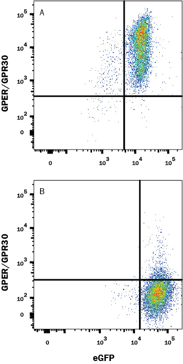
Detection of GPER/GPR30 in HEK293 Human Cell Line Transfected with human GPER/GRP30 and eGFP by Flow Cytometry. HEK293 human embryonic kidney cell line transfected with (A) human GPER/GRP30 or (B) irrelevant transfectants, and eGFP was stained with Goat Anti-Human GPER/GPR30 Affinity-Purified Polyclonal Antibody (Catalog # AF5534) followed by Allophycocyanin-conjugated Anti-Goat IgG Secondary Antibody (Catalog # F0108). Quadrants were set based on Goat IgG Flow Cytometry Isotype Control (Catalog # AB-108-C, data not shown). View our protocol for Staining Membrane-associated Proteins.
 View Larger
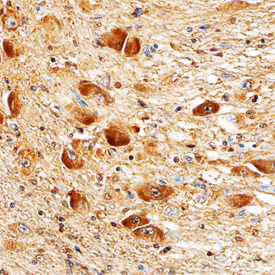
GPER in Human Brain. GPER was detected in immersion fixed paraffin-embedded sections of human brain (hypothalamus) using Goat Anti-Human GPER Antigen Affinity-purified Polyclonal Antibody (Catalog # AF5534) at 10 µg/mL overnight at 4 °C. Tissue was stained using the Anti-Goat HRP-DAB Cell & Tissue Staining Kit (brown; Catalog # CTS008) and counterstained with hematoxylin (blue). Specific staining was localized to neuronal cell bodies. View our protocol for Chromogenic IHC Staining of Paraffin-embedded Tissue Sections.
Reconstitution Calculator
Preparation and Storage
- 12 months from date of receipt, -20 to -70 °C as supplied.
- 1 month, 2 to 8 °C under sterile conditions after reconstitution.
- 6 months, -20 to -70 °C under sterile conditions after reconstitution.
Background: GPER/GPR30
GPER (G-Protein Coupled Estrogen Receptor 1; also GPR30, DRY12 and mER) is a 44 kDa, seven transmembrane (TM) member of the GPR-1 family of molecules. It is ubiquitously expressed, appearing on/in neurons, monocytes and endothelial cells. Its exact location is unclear; it has been described in both the cell membrane and ER, but not by all investigators. Human GPER is 375 amino acids (aa) in length. It contains an N-terminal extracellular region (aa 1‑62), a series of seven TM domains (aa 63‑327), and a C-terminal cytoplasmic tail (aa 328‑375). The initial function attributed to GPER was that of a membrane receptor for estrogen. This is in dispute. There are two potential splice variants for GPER. One shows a deletion of aa 32-49, while a second shows a 99 aa substitution for aa 308‑375. Over aa 1‑62, human GPER shares 57% aa identity with mouse GPER.
Product Datasheets
Citations for Human GPER/GPR30 Antibody
R&D Systems personnel manually curate a database that contains references using R&D Systems products. The data collected includes not only links to publications in PubMed, but also provides information about sample types, species, and experimental conditions.
4
Citations: Showing 1 - 4
Filter your results:
Filter by:
-
UDP-6-glucose dehydrogenase in hormonally responsive breast cancers
Authors: Price, MJ;Nguyen, AD;Haines, C;Baëta, CD;Byemerwa, J;Murkajee, D;Artham, S;Kumar, V;Lavau, C;Wardell, S;Varghese, S;Goodwin, CR;
bioRxiv : the preprint server for biology
Species: Mouse
Sample Types: Protein
Applications: Western Blot -
Assessment of G Protein-Coupled Oestrogen Receptor Expression in Normal and Neoplastic Human Tissues Using a Novel Rabbit Monoclonal Antibody
Authors: M Bubb, AL Beyer, P Dasgupta, D Kaemmerer, J Sänger, K Evert, RM Wirtz, S Schulz, A Lupp
International Journal of Molecular Sciences, 2022-05-06;23(9):.
Species: Human
Sample Types: Whole Tissue
Applications: IHC -
Plasma membrane expression of G protein-coupled estrogen receptor (GPER)/G protein-coupled receptor 30 (GPR30) is associated with worse outcome in metachronous contralateral breast cancer
Authors: J Tutzauer, M Sjöström, PO Bendahl, L Rydén, M Fernö, LMF Leeb-Lundb, S Alkner
PLoS ONE, 2020-04-17;15(4):e0231786.
Species: Human
Sample Types: Cell Lysates
-
G protein-coupled receptor 30 (GPR30) forms a plasma membrane complex with membrane-associated guanylate kinases (MAGUKs) and protein kinase A-anchoring protein 5 (AKAP5) that constitutively inhibits cAMP production.
Authors: Broselid S, Berg K, Chavera T, Kahn R, Clarke W, Olde B, Leeb-Lundberg L
J Biol Chem, 2014-06-24;289(32):22117-27.
Species: Human
Sample Types: Whole Cells
Applications: IHC-Fr, Western Blot
FAQs
No product specific FAQs exist for this product, however you may
View all Antibody FAQsReviews for Human GPER/GPR30 Antibody
There are currently no reviews for this product. Be the first to review Human GPER/GPR30 Antibody and earn rewards!
Have you used Human GPER/GPR30 Antibody?
Submit a review and receive an Amazon gift card.
$25/€18/£15/$25CAN/¥75 Yuan/¥2500 Yen for a review with an image
$10/€7/£6/$10 CAD/¥70 Yuan/¥1110 Yen for a review without an image

