Human HGFR/c-MET Antibody Summary
Glu25-Thr932
Accession # P08581
Applications
Please Note: Optimal dilutions should be determined by each laboratory for each application. General Protocols are available in the Technical Information section on our website.
Scientific Data
.") View Larger
View Larger
HGF R/c‑MET in HT‑29 and U937 Human Cell Line. HGF R/c-MET was detected in immersion fixed HT-29 human colon adenocarcinoma cell line (positive control, left panel) and U937 human histiocytic lymphoma cell line (negative control, right panel) using Goat Anti-Human HGF R/c-MET Antigen Affinity-purified Polyclonal Antibody (Catalog # AF276) at 5 µg/mL for 3 hours at room temperature. Cells were stained using the NorthernLights™ 557-conjugated Anti-Goat IgG Secondary Antibody (red; NL001) and counterstained with DAPI (blue). Specific staining was localized to plasma membrane. View our protocol for Fluorescent ICC Staining of Cells on Coverslips.
.") View Larger
View Larger
HGF R/c‑MET in Human Liver. HGF R/c-MET was detected in immersion fixed paraffin-embedded sections of human liver using Goat Anti-Human HGF R/c-MET Antigen Affinity-purified Polyclonal Antibody (Catalog # AF276) at 10 µg/mL overnight at 4 °C. Before incubation with the primary antibody tissue was subjected to heat-induced epitope retrieval using Antigen Retrieval Reagent-Basic (CTS013). Tissue was stained using the Anti-Goat HRP-DAB Cell & Tissue Staining Kit (brown; CTS008) and counterstained with hematoxylin (blue). View our protocol for Chromogenic IHC Staining of Paraffin-embedded Tissue Sections.
.") View Larger
View Larger
HGF R/c‑MET in Human Skin. HGF R/c-MET was detected in immersion fixed paraffin-embedded sections of human skin using 15 µg/mL Goat Anti-Human HGF R/c-MET Antigen Affinity-purified Polyclonal Antibody (Catalog # AF276) overnight at 4 °C. Tissue was stained with the Anti-Goat HRP-DAB Cell & Tissue Staining Kit (brown; CTS008) and counterstained with hematoxylin (blue). View our protocol for Chromogenic IHC Staining of Paraffin-embedded Tissue Sections.
 View Larger
Western Blot Shows Human HGF R/c‑MET Specificity by Using Knockout Cell Line. Western blot shows lysates of HeLa human cervical epithelial carcinoma parental cell line and HGF R/c-Met knockout HeLa cell line (KO). PVDF membrane was probed with 1 µg/mL of Goat Anti-Human HGF R/c-MET Antigen Affinity-purified Polyclonal Antibody (Catalog # AF276) followed by HRP-conjugated Anti-Goat IgG Secondary Antibody (HAF017). Specific bands were detected for HGF R/c-MET at approximately 150-200 kDa (as indicated) in the parental HeLa cell line, but is not detectable in knockout HeLa cell line. GAPDH (AF5718) is shown as a loading control. This experiment was conducted under reducing conditions and using Immunoblot Buffer Group 1.
 View Larger
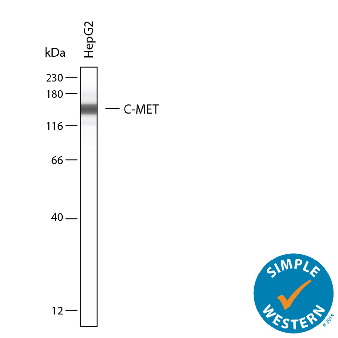
Detection of Human HGFR/c-MET by Simple WesternTM. Simple Western lane view shows lysates of HepG2 human hepatocellular carcinoma cell line, loaded at 0.2 mg/mL. A specific band was detected for HGFR/c-MET at approximately 151 kDa (as indicated) using 25 µg/mL of Goat Anti-Human HGFR/c-MET Antigen Affinity-purified Polyclonal Antibody (Catalog # AF276). This experiment was conducted under reducing conditions and using the 12-230 kDa separation system.
Reconstitution Calculator
Preparation and Storage
- 12 months from date of receipt, -20 to -70 °C as supplied.
- 1 month, 2 to 8 °C under sterile conditions after reconstitution.
- 6 months, -20 to -70 °C under sterile conditions after reconstitution.
Background: HGFR/c-MET
HGF R, also known as Met (from N-methyl-N’-nitro-N-nitrosoguanidine induced), is a glycosylated receptor tyrosine kinase that plays a central role in epithelial morphogenesis and cancer development. HGF R is synthesized as a single chain precursor which undergoes cotranslational proteolytic cleavage. This generates a mature HGF R that is a disulfide-linked dimer composed of a 50 kDa extracellular alpha chain and a 145 kDa transmembrane beta chain (1, 2). The extracellular domain (ECD) contains a seven bladed beta -propeller sema domain, a cysteine-rich PSI/MRS, and four Ig-like E-set domains, while the cytoplasmic region includes the tyrosine kinase domain (3, 4). Proteolysis and alternate splicing generate additional forms of human HGF R which either lack of the kinase domain, consist of secreted extracellular domains, or are deficient in proteolytic separation of the alpha and beta chains (5-7). The sema domain, which is formed by both the alpha and beta chains of HGF R, mediates both ligand binding and receptor dimerization (3, 8). Ligand-induced tyrosine phosphorylation in the cytoplasmic region activates the kinase domain and provides docking sites for multiple SH2-containing molecules (9, 10). HGF stimulation induces HGF R downregulation via internalization and proteasome-dependent degradation (11). In the absence of ligand, HGF R forms non-covalent complexes with a variety of membrane proteins including CD44v6, CD151, EGF R, Fas, Integrin alpha 6/ beta 4, Plexins B1, 2, 3, and MSP R/Ron (12-19). Ligation of one complex component triggers activation of the other, followed by cooperative signaling effects (12-19). Formation of some of these heteromeric complexes is a requirement for epithelial cell morphogenesis and tumor cell invasion (12, 16, 17). Paracrine induction of epithelial cell scattering and branching tubulogenesis results from the stimulation of HGF R on undifferentiated epithelium by HGF released from neighboring mesenchymal cells (20). Genetic polymorphisms, chromosomal translocation, overexpression, and additional splicing and proteolytic cleavage of HGF R have been described in a wide range of cancers (1). Within the ECD, human HGF R shares 86-88% aa sequence identity with canine, mouse, and rat HGF R.
- Birchmeier, C. et al. (2003) Nat. Rev. Mol. Cell Biol. 4:915.
- Corso, S. et al. (2005) Trends Mol. Med. 11:284.
- Gherardi, E. et al. (2003) Proc. Natl. Acad. Sci. USA 100:12039.
- Park, M. et al. (1987) Proc. Natl. Acad. Sci. USA 84:6379.
- Crepaldi, T. et al. (1994) J. Biol. Chem. 269:1750.
- Prat, M. et al. (1991) Mol. Cell. Biol. 12:5954.
- Rodrigues, G.A. et al. (1991) Mol. Cell. Biol. 11:2962.
- Kong-Beltran, M. et al. (2004) Cancer Cell 6:75.
- Naldini, L. et al. (1991) Mol. Cell. Biol. 11:1793.
- Ponzetto, C. et al. (1994) Cell 77:261.
- Jeffers, M. et al. (1997) Mol. Cell. Biol. 17:799.
- Orian-Rousseau, V. et al. (2002) Genes Dev. 16:3074.
- Klosek, S.K. et al. (2005) Biochem. Biophys. Res. Commun. 336:408.
- Jo, M. et al. (2000) J. Biol. Chem. 275:8806.
- Wang, X. et al. (2002) Mol. Cell 9:411.
- Trusolino, L. et al. (2001) Cell 107:643.
- Giordano, S. et al. (2002) Nat. Cell Biol. 4:720.
- Conrotto, P. et al. (2004) Oncogene 23:5131.
- Follenzi, A. et al. (2000) Oncogene 19:3041.
- Sonnenberg, E. et al. (1993) J. Cell Biol. 123:223.
Product Datasheets
Citations for Human HGFR/c-MET Antibody
R&D Systems personnel manually curate a database that contains references using R&D Systems products. The data collected includes not only links to publications in PubMed, but also provides information about sample types, species, and experimental conditions.
26
Citations: Showing 1 - 10
Filter your results:
Filter by:
-
Expression and Prognostic Evaluation of the Receptor Tyrosine Kinase MET in Canine Malignant Melanoma
Authors: K Koo, A Wuenschman, A Rendahl, KY Song, C Forster, A Wolf-Ringw, A Borgatti, A Giubellino
Veterinary sciences, 2023-03-26;10(4):.
Species: Canine
Sample Types: Cell Lysates
Applications: Western Blot -
Plexin-B3 expression stimulates MET signaling, breast cancer stem cell specification, and lung metastasis.
Authors: Zuo Q, Yang Y, Lyu Y, Yang C, Chen C, Salman S, Huang T, Wicks E, Jackson W, Datan E, Qin W, Semenza G
Cell Rep, 2023-02-28;42(3):112164.
Species: Human
Sample Types: Cell Lysates
Applications: Western Blot -
The Impact of Nanobody Density on the Targeting Efficiency of PEGylated Liposomes
Authors: BS Mesquita, MHAM Fens, A Di Maggio, EDC Bosman, WE Hennink, M Heger, S Oliveira
International Journal of Molecular Sciences, 2022-11-29;23(23):.
Species: Human
Sample Types: Whole Cells
Applications: Flow Cytometry -
Supermeres are functional extracellular nanoparticles replete with disease biomarkers and therapeutic targets
Authors: Q Zhang, DK Jeppesen, JN Higginboth, R Graves-Dea, VQ Trinh, MA Ramirez, Y Sohn, AC Neininger, N Taneja, ET McKinley, H Niitsu, Z Cao, R Evans, SE Glass, KC Ray, WH Fissell, S Hill, KL Rose, WJ Huh, MK Washington, GD Ayers, DT Burnette, S Sharma, LH Rome, JL Franklin, YA Lee, Q Liu, RJ Coffey
Nature Cell Biology, 2021-12-09;23(12):1240-1254.
Species: Human
Sample Types: Cell Lysates
Applications: Western Blot -
The EMT activator ZEB1 accelerates endosomal trafficking to establish a polarity axis in lung adenocarcinoma cells
Authors: P Banerjee, GY Xiao, X Tan, VJ Zheng, L Shi, MNB Rabassedas, HF Guo, X Liu, J Yu, L Diao, J Wang, WK Russell, J Roszik, CJ Creighton, JM Kurie
Nature Communications, 2021-11-03;12(1):6354.
Species: Human
Sample Types: Whole Cells
Applications: ICC -
RAL GTPases mediate EGFR-driven intestinal stem cell proliferation and tumourigenesis
Authors: M Nászai, K Bellec, Y Yu, A Román-Fern, E Sandilands, J Johansson, AD Campbell, JC Norman, OJ Sansom, DM Bryant, JB Cordero
Elife, 2021-06-07;10(0):.
Species: Human
Sample Types: Cell Lysates
Applications: ELISA Capture -
The Role of Receptor Tyrosine Kinases in Lassa Virus Cell Entry
Authors: C Fedeli, H Moreno, S Kunz
Viruses, 2020-08-06;12(8):.
Species: Human
Sample Types: Whole Cells
Applications: Flow Cytometry -
FCHSD2 controls oncogenic ERK1/2 signaling outcome by regulating endocytic trafficking
Authors: GY Xiao, SL Schmid
PLoS Biol., 2020-07-17;18(7):e3000778.
Species: Human
Sample Types: Whole Cells
Applications: Flow Cytometry -
Broad-spectrum receptor tyrosine kinase inhibitors overcome de novo and acquired modes of resistance to EGFR-targeted therapies in colorectal cancer
Authors: R Graves-Dea, G Bogatcheva, S Rehman, Y Lu, JN Higginboth, B Singh
Oncotarget, 2019-02-12;10(13):1320-1333.
Species: Human
Sample Types: Cell Lysates
Applications: Western Blot -
Mutant p53s generate pro-invasive niches by influencing exosome podocalyxin levels
Authors: D Novo, N Heath, L Mitchell, G Caligiuri, A MacFarlane, D Reijmer, L Charlton, J Knight, M Calka, E McGhee, E Dornier, D Sumpton, S Mason, A Echard, K Klinkert, J Secklehner, F Kruiswijk, K Vousden, IR Macpherson, K Blyth, P Bailey, H Yin, LM Carlin, J Morton, S Zanivan, JC Norman
Nat Commun, 2018-11-29;9(1):5069.
Species: Human
Sample Types: Whole Cells
Applications: Bioassay -
The association of semaphorin 5A with lymph node metastasis and adverse prognosis in cervical cancer
Authors: JB Xiao, XL Li, L Liu, G Wang, SN Hao, HJ Dong, XM Wang, YF Zhang, HD Liu
Cancer Cell Int., 2018-06-22;18(0):87.
Species: Human
Sample Types: Whole Cells
Applications: Neutralization -
Human papillomavirus type 16 E5-mediated upregulation of Met in human keratinocytes
Authors: ML Scott, DT Coleman, KC Kelly, JL Carroll, B Woodby, WK Songock, JA Cardelli, JM Bodily
Virology, 2018-03-31;519(0):1-11.
Species: Human
Sample Types: Whole Cells
Applications: ICC -
Beta 1-integrin-c-Met cooperation reveals an inside-in survival signalling on autophagy-related endomembranes
Authors: Rachel Barrow-McG
Nat Commun, 2016-06-23;7(0):11942.
Species: Human
Sample Types: Cell Lysates
Applications: Western Blot -
Tumor and Plasma Met Levels in Non-Metastatic Prostate Cancer
Authors: Deborah R Kaye
PLoS ONE, 2016-06-14;11(6):e0157130.
Species: Human
Sample Types: Plasma
Applications: ELISA Development (Detection) -
Targeting matriptase in breast cancer abrogates tumour progression via impairment of stromal-epithelial growth factor signalling.
Authors: Zoratti G, Tanabe L, Varela F, Murray A, Bergum C, Colombo E, Lang J, Molinolo A, Leduc R, Marsault E, Boerner J, List K
Nat Commun, 2015-04-15;6(0):6776.
Species: Human
Sample Types: Whole Tissue
Applications: IHC-P -
A pharmacodynamic/pharmacokinetic study of ficlatuzumab in patients with advanced solid tumors and liver metastases.
Authors: Tabernero J, Elez M, Herranz M, Rico I, Prudkin L, Andreu J, Mateos J, Carreras M, Han M, Gifford J, Credi M, Yin W, Agarwal S, Komarnitsky P, Baselga J
Clin Cancer Res, 2014-03-14;20(10):2793-804.
Species: Human
Sample Types: Serum
Applications: ELISA Development (Detection) -
Internalization of Met requires the co-receptor CD44v6 and its link to ERM proteins.
Authors: Hasenauer S, Malinger D, Koschut D, Pace G, Matzke A, von Au A, Orian-Rousseau V
PLoS ONE, 2013-04-23;8(4):e62357.
Species: Human
Sample Types: Whole Cells
Applications: ICC -
Mutant p53 enhances MET trafficking and signalling to drive cell scattering and invasion.
Authors: Muller, P A J, Trinidad, A G, Timpson, P, Morton, J P, Zanivan, S, van den Berghe, P V E, Nixon, C, Karim, S A, Caswell, P T, Noll, J E, Coffill, C R, Lane, D P, Sansom, O J, Neilsen, P M, Norman, J C, Vousden, K H
Oncogene, 2012-05-14;32(10):1252-65.
Species: Human
Sample Types: Cell Lysates
Applications: Western Blot -
Identification of a pivotal endocytosis motif in c-Met and selective modulation of HGF-dependent aggressiveness of cancer using the 16-mer endocytic peptide.
Authors: Cho K, Park J, Park C, Lee D, Lee E, Kim D, Kim K, Yoon S, Park Y, Kim E, Cho S, Jang S, Park B, Chi S, Yoo S, Jang M, Kim H, Kim E, Jo K, Park Y
Oncogene, 2012-04-23;32(8):1018-29.
Species: Human
Sample Types: Whole Cells
Applications: Flow Cytometry -
c-Met-induced epithelial carcinogenesis is initiated by the serine protease matriptase.
Authors: Szabo R, Rasmussen AL, Moyer AB
Oncogene, 2011-01-10;30(17):2003-16.
Species: Human
Sample Types: Whole Tissue
Applications: IHC-P -
Dorsal ruffle microdomains potentiate Met receptor tyrosine kinase signaling and down-regulation.
Authors: Abella JV, Parachoniak CA, Sangwan V, Park M
J. Biol. Chem., 2010-06-07;285(32):24956-67.
Species: Human
Sample Types: Whole Cells
Applications: ICC -
Epidermal growth factor receptor regulates MET levels and invasiveness through hypoxia-inducible factor-1alpha in non-small cell lung cancer cells.
Authors: Xu L, Nilsson MB, Saintigny P, Cascone T, Herynk MH, Du Z, Nikolinakos PG, Yang Y, Prudkin L, Liu D, Lee JJ, Johnson FM, Wong KK, Girard L, Gazdar AF, Minna JD, Kurie JM, Wistuba II, Heymach JV
Oncogene, 2010-02-15;29(18):2616-27.
Species: Human
Sample Types: Whole Tissue
Applications: IHC-P -
Effect of human patient plasma ex vivo treatment on gene expression and progenitor cell activation of primary human liver cells in multi-compartment 3D perfusion bioreactors for extra-corporeal liver support.
Authors: Schmelzer E, Mutig K, Schrade P, Bachmann S, Gerlach JC, Zeilinger K
Biotechnol. Bioeng., 2009-07-01;103(4):817-27.
Species: Human
Sample Types: Whole Tissue
Applications: IHC-P -
Decorin is a novel antagonistic ligand of the Met receptor.
Authors: Goldoni S, Humphries A, Nystrom A, Sattar S, Owens RT, McQuillan DJ, Ireton K, Iozzo RV
J. Cell Biol., 2009-05-11;185(4):743-54.
Species: Human
Sample Types: Whole Cells
Applications: ICC -
Impairment of the antifibrotic effect of hepatocyte growth factor in lung fibroblasts from African Americans: possible role in systemic sclerosis.
Authors: Bogatkevich GS, Ludwicka-Bradley A, Highland KB, Hant F, Nietert PJ, Singleton CB, Feghali-Bostwick CA, Silver RM
Arthritis Rheum., 2007-07-01;56(7):2432-42.
Species: Human
Sample Types: Whole Cells
Applications: Neutralization -
Bronchial epithelial cell growth regulation in fibroblast cocultures: the role of hepatocyte growth factor.
Authors: Skibinski G, Elborn JS, Ennis M
Am. J. Physiol. Lung Cell Mol. Physiol., 2007-03-23;293(1):L69-76.
Species: Human
Sample Types: Whole Cells
Applications: Neutralization
FAQs
No product specific FAQs exist for this product, however you may
View all Antibody FAQsReviews for Human HGFR/c-MET Antibody
There are currently no reviews for this product. Be the first to review Human HGFR/c-MET Antibody and earn rewards!
Have you used Human HGFR/c-MET Antibody?
Submit a review and receive an Amazon gift card.
$25/€18/£15/$25CAN/¥75 Yuan/¥2500 Yen for a review with an image
$10/€7/£6/$10 CAD/¥70 Yuan/¥1110 Yen for a review without an image


