Human IGF-II R/IGF2R Antibody Summary
Ser1510-Phe2108
Accession # P11717
Applications
Please Note: Optimal dilutions should be determined by each laboratory for each application. General Protocols are available in the Technical Information section on our website.
Scientific Data
 View Larger
Detection of IGF-II R/IGF2R in Human Monocytes by Flow Cytometry. Human whole blood monocytes were stained with Goat Anti-Human IGF-II R/IGF2R Antigen Affinity-purified Polyclonal Antibody (Catalog # AF2447, filled histogram) or control antibody (Catalog # AB-108-C, open histogram), followed by Phycoerythrin-conjugated Anti-Goat IgG Secondary Antibody (Catalog # F0107).
 View Larger
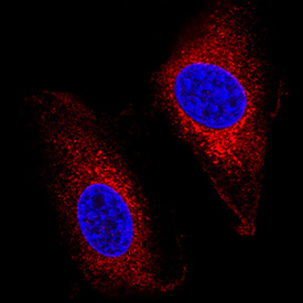
IGF-II R/IGF2R in A172 Human Cell Line. IGF-II R/IGF2R was detected in immersion fixed A172 human glioblastoma cell line using Goat Anti-Human IGF-II R/IGF2R Antigen Affinity-purified Polyclonal Antibody (Catalog # AF2447) at 5 µg/mL for 3 hours at room temperature. Cells were stained using the NorthernLights™ 557-conjugated Anti-Goat IgG Secondary Antibody (red; Catalog # NL001) and counterstained with DAPI (blue). Specific staining was localized to cytoplasm. View our protocol for Fluorescent ICC Staining of Cells on Coverslips.
 View Larger
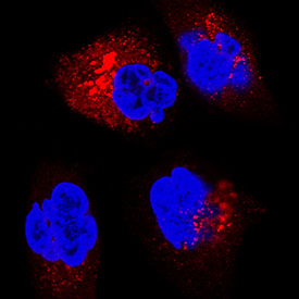
IGF-II R/IGF2R in A549 Human Cell Line. IGF-II R/IGF2R was detected in immersion fixed A549 human lung carcinoma cell line using Goat Anti-Human IGF-II R/IGF2R Antigen Affinity-purified Polyclonal Antibody (Catalog # AF2447) at 5 µg/mL for 3 hours at room temperature. Cells were stained using the NorthernLights™ 557-conjugated Anti-Goat IgG Secondary Antibody (red; Catalog # NL001) and counterstained with DAPI (blue). Specific staining was localized to cell surface and cytoplasm. View our protocol for Fluorescent ICC Staining of Cells on Coverslips.
 View Larger
IGF-II R/IGF2R R in Human Placenta. IGF-II R/IGF2R was detected in immersion fixed paraffin-embedded sections of human placenta using 10 µg/mL Goat Anti-Human IGF-II R/IGF2R Antigen Affinity-purified Polyclonal Antibody (Catalog # AF2447) overnight at 4 °C. Tissue was stained with the Anti-Goat HRP-DAB Cell & Tissue Staining Kit (brown; Catalog # CTS008) and counterstained with hematoxylin (blue). View our protocol for Chromogenic IHC Staining of Paraffin-embedded Tissue Sections.
Reconstitution Calculator
Preparation and Storage
- 12 months from date of receipt, -20 to -70 °C as supplied.
- 1 month, 2 to 8 °C under sterile conditions after reconstitution.
- 6 months, -20 to -70 °C under sterile conditions after reconstitution.
Background: IGF-II R/IGF2R
The type 2 insulin-like growth factor receptor (also known as cation-independent mannose-6 phosphate receptor/CI-MPR) is a 300 kDa member of the P-type lectin family of molecules. P-type lectins generate functional eukaryotic lysosomes by binding and sorting lysosomal enzymes expressing phosphorylated mannose residues (M6P) (1-3). IGF-II R is a type I transmembrane glycoprotein that contains a 2,264 amino acid (aa) extracellular region, a 23 aa transmembrane segment and a 124 aa cytoplasmic tail (4, 5). The extracellular region consists of 15 contiguous “binding” repeats of about 150 aa each. The odd-numbered repeats interact with “ligands” while the even-numbered repeats likely generate a nondisulfide homodimer in the membrane (1). Repeat #11 binds IGF-II, while repeats 3 and 9 bind mannose-6 phosphate; repeat #13 contains a fibronectin type II motif and assists in IGF-II binding (1, 2). In the extracellular region of IGF-II R expressed by R&D Systems (600 aa’s), human IGF-II R is 85% aa identical to both mouse and bovine IGF-II R. This expressed region includes binding repeats #11, 12, and 13. In addition to IGF-II, CI-MPR/IGF-II R binds non-M6P containing ligands such as retinoic acid, urokinase-type plasminogen-activator receptor and plasminogen, plus M6P‑containing molecules such as lysosomal enzymes, TGF-beta 1 precursor, proliferin, LIF, CD26, herpes simplex glycoprotein D, and granzymes A and B (2, 6). IGF-II R regulates many diverse biological functions that range from intracellular trafficking to the internalization of extracellular factors and modulation of cellular responses. It delivers newly synthesized M6P-tagged lysosomal enzymes from the trans-golgi network to endosomes, and facilitates the clearance of extracellular lysosomal and matrix degrading enzymes by internalization into clathrin-coated vesicles and delivery into endosomes. With respect to IGF-II biology, it would appear that IGF-II R is principally a regulator of local IGF-II levels, targeting IGF-II for destruction in lysosomes (2). However, some evidence suggests the receptor will signal via G‑proteins, an effect that has yet to be conclusively shown (6).
- Ghosh, P. et al. (2003) Nat. Rev. Mol. Cell. Biol. 4:202.
- Dahms, N.M. and M.K. Hancock (2002) Biochim. Biophys. Acta. 1572:317.
- Zaina, S. and J. Nilsson (2003) Curr. Opin. Lipidol. 14:483.
- Morgan, D.O. et al. (1987) Nature 329:301.
- Oshima, A. et al. (1988) J. Biol. Chem. 263:2553.
- Hawkes, C. and S. Kar (2004) Brain Res. Rev. 44:117.
Product Datasheets
Citations for Human IGF-II R/IGF2R Antibody
R&D Systems personnel manually curate a database that contains references using R&D Systems products. The data collected includes not only links to publications in PubMed, but also provides information about sample types, species, and experimental conditions.
10
Citations: Showing 1 - 10
Filter your results:
Filter by:
-
Blockade of IGF2R improves muscle regeneration and ameliorates Duchenne muscular dystrophy
Authors: P Bella, A Farini, S Banfi, D Parolini, N Tonna, M Meregalli, M Belicchi, S Erratico, P D'Ursi, F Bianco, M Legato, C Ruocco, C Sitzia, S Sangiorgi, C Villa, G D'Antona, L Milanesi, E Nisoli, P Mauri, Y Torrente
EMBO Mol Med, 2019-12-02;0(0):e11019.
Species: Mouse
Sample Types: In Vivo
Applications: In Vivo -
Insulin stimulates PI3K/AKT and cell adhesion to promote the survival of individualized human embryonic stem cells
Authors: C Godoy-Pare, C Deng, W Liu, G Chen
Stem Cells, 2019-05-17;0(0):.
Species: Human
Sample Types: Cell Lysates
Applications: Western Blot -
The APMAP interactome reveals new modulators of APP processing and beta-amyloid production that are altered in Alzheimer's disease
Authors: H Gerber, S Mosser, B Boury-Jamo, M Stumpe, A Piersigill, C Goepfert, J Dengjel, U Albrecht, F Magara, PC Fraering
Acta Neuropathol Commun, 2019-01-31;7(1):13.
Species: Mouse
Sample Types: Tissue Homogenates
Applications: Western Blot -
IGF-II promotes neuroprotection and neuroplasticity recovery in a long-lasting model of oxidative damage induced by glucocorticoids
Authors: E Martín-Mon, C Millon, F Boraldi, F Garcia-Gui, C Pedraza, E Lara, LJ Santin, J Pavia, M Garcia-Fer
Redox Biol, 2017-05-26;13(0):69-81.
Species: Rat
Sample Types: Whole Cells
Applications: Neutralization -
mTORC1 regulates mannose-6-phosphate receptor transport and T-cell vulnerability to regulatory T cells by controlling kinesin KIF13A
Authors: KA Ahmed, J Xiang
Cell Discov, 2017-04-25;3(0):17011.
Species: Mouse
Sample Types: Whole Cells
Applications: Flow Cytometry -
A subset of bone marrow stromal cells regulate ATP-binding cassette gene expression via insulin-like growth factor-I in a leukemia cell line.
Authors: Benabbou N, Mirshahi P, Bordu C, Faussat A, Tang R, Therwath A, Soria J, Marie J, Mirshahi M
Int J Oncol, 2014-07-29;45(4):1372-80.
Species: Human
Sample Types: Whole Cells
Applications: ICC -
A critical role for IGF-II in memory consolidation and enhancement.
Authors: Chen DY, Stern SA, Garcia-Osta A, Saunier-Rebori B, Pollonini G, Bambah-Mukku D, Blitzer RD, Alberini CM
Nature, 2011-01-27;469(7331):491-7.
Species: Rat
Sample Types: In Vivo
Applications: Neutralization -
IGF2 actions on trophoblast in human placenta are regulated by the insulin-like growth factor 2 receptor, which can function as both a signaling and clearance receptor.
Authors: Harris LK, Crocker IP, Baker PN, Aplin JD, Westwood M
Biol. Reprod., 2010-10-27;84(3):440-6.
Species: Human
Sample Types: Cell Lysates, Whole Cells
Applications: ICC, Western Blot -
CREG inhibits migration of human vascular smooth muscle cells by mediating IGF-II endocytosis.
Authors: Han Y, Cui J, Tao J, Guo L, Guo P, Sun M, Kang J, Zhang X, Yan C, Li S
Exp. Cell Res., 2009-09-19;315(19):3301-11.
Species: Human
Sample Types: Cell Lysates, Whole Cells
Applications: ICC, Neutralization, Western Blot -
Urokinase induces survival or pro-apoptotic signals in human mesangial cells depending on the apoptotic stimulus.
Authors: Tkachuk N, Kiyan J, Tkachuk S, Kiyan R, Shushakova N, Haller H, Dumler I
Biochem. J., 2008-10-15;415(2):265-73.
Species: Human
Sample Types: Whole Cells
Applications: ICC
FAQs
No product specific FAQs exist for this product, however you may
View all Antibody FAQsReviews for Human IGF-II R/IGF2R Antibody
There are currently no reviews for this product. Be the first to review Human IGF-II R/IGF2R Antibody and earn rewards!
Have you used Human IGF-II R/IGF2R Antibody?
Submit a review and receive an Amazon gift card.
$25/€18/£15/$25CAN/¥75 Yuan/¥2500 Yen for a review with an image
$10/€7/£6/$10 CAD/¥70 Yuan/¥1110 Yen for a review without an image


