Human IL-3 Quantikine ELISA Kit Summary
Product Summary
Precision
Cell Culture Supernates
| Intra-Assay Precision | Inter-Assay Precision | |||||
|---|---|---|---|---|---|---|
| Sample | 1 | 2 | 3 | 1 | 2 | 3 |
| n | 20 | 20 | 20 | 20 | 20 | 20 |
| Mean (pg/mL) | 126 | 731 | 991 | 128 | 725 | 1423 |
| Standard Deviation | 5.7 | 23.7 | 30.4 | 10.1 | 45.9 | 81.5 |
| CV% | 4.5 | 3.2 | 3.1 | 7.9 | 6.3 | 5.7 |
Serum, EDTA Plasma, Heparin Plasma, Citrate Plasma
| Intra-Assay Precision | Inter-Assay Precision | |||||
|---|---|---|---|---|---|---|
| Sample | 1 | 2 | 3 | 1 | 2 | 3 |
| n | 20 | 20 | 20 | 20 | 20 | 20 |
| Mean (pg/mL) | 115 | 728 | 1460 | 124 | 737 | 1450 |
| Standard Deviation | 6.5 | 23.6 | 48.7 | 13.3 | 41.4 | 104 |
| CV% | 5.7 | 3.2 | 3.3 | 10.7 | 5.6 | 7.2 |
Recovery
The recovery of human IL-3 spiked to levels throughout the range of the assay in various matrices was evaluated.
| Sample Type | Average % Recovery | Range % |
|---|---|---|
| Cell Culture Media (n=5) | 92 | 78-105 |
| Citrate Plasma (n=) | 104 | 93-122 |
| EDTA Plasma (n=) | 108 | 96-117 |
| Heparin Plasma (n=) | 100 | 93-108 |
| Serum (n=5) | 104 | 96-118 |
Linearity
Scientific Data
Product Datasheets
Preparation and Storage
Background: IL-3
M-CSF, also known as CSF-1, is a four-alpha-helical-bundle cytokine that is the primary regulator of macrophage survival, proliferation and differentiation. M-CSF is also essential for the survival and proliferation of osteoclast progenitors. M-CSF also primes and enhances macrophage killing of tumor cells and microorganisms, regulates the release of cytokines and other inflammatory modulators from macrophages, and stimulates pinocytosis. M-CSF increases during pregnancy to support implantation and growth of the decidua and placenta. Sources of M-CSF include fibroblasts, activated macrophages, endometrial secretory epithelium, bone marrow stromal cells and activated endothelial cells. The M-CSF receptor (c-fms) transduces its pleotropic effects and mediates its endocytosis. M-CSF mRNAs of various sizes occur. Full length human M-CSF transcripts encode a 522 amino acid (aa) type I transmembrane (TM) protein with a 464 aa extracellular region, a 21 aa TM domain, and a 37 aa cytoplasmic tail that forms a 140 kDa covalent dimer. Differential processing produces two proteolytically cleaved, secreted dimers. One is an N- and O- glycosylated 86 kDa dimer, while the other is modified by both glycosylation and chondroitin-sulfate proteoglycan (PG) to generate a 200 kDa subunit. Although PG-modified M-CSF can circulate, it may be immobilized by attachment to type V collagen. Shorter transcripts encode M-CSF that lack cleavage and PG sites and produce an N-glycosylated 68 kDa TM dimer and a slowly produced 44 kDa secreted dimer. Although forms may vary in activity and half-life, all contain the N-terminal 150 aa portion that is necessary and sufficient for interaction with the M-CSF receptor. The first 223 aa of mature human M-CSF shares 88%, 86%, 81% and 74% aa identity with corresponding regions of dog, cow, mouse and rat M-CSF, respectively. Human M-CSF is active in the mouse, but mouse M-CSF is reported to be species-specific.
Assay Procedure
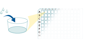
Refer to the product- Prepare all reagents, standard dilutions, and samples as directed in the product insert.
- Remove excess microplate strips from the plate frame, return them to the foil pouch containing the desiccant pack, and reseal.
- For Serum & Plasma Samples Only: Add 50 µL of Assay Diluent to each well.
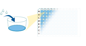
- Add 200 µL of Standard, control, or sample to each well. Cover with a plate sealer, and incubate at room temperature for 2 hours.
- Aspirate each well and wash, repeating the process 3 times for a total of 4 washes.
- Add 200 µL of Conjugate to each well. Cover with a new plate sealer, and incubate at room temperature for 2 hours.
- Aspirate and wash 4 times.
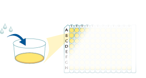
- Add 200 µL Substrate Solution to each well. Incubate at room temperature for 20 minutes. PROTECT FROM LIGHT.
- Add 50 µL of Stop Solution to each well. Read at 450 nm within 30 minutes. Set wavelength correction to 540 nm or 570 nm.


Citations for Human IL-3 Quantikine ELISA Kit
R&D Systems personnel manually curate a database that contains references using R&D Systems products. The data collected includes not only links to publications in PubMed, but also provides information about sample types, species, and experimental conditions.
17
Citations: Showing 1 - 10
Filter your results:
Filter by:
-
Inflammatory biomarkers, geriatric assessment, and treatment outcomes in acute myeloid leukemia
Authors: KP Loh, JA Tooze, BJ Nicklas, SB Kritchevsk, JD Williamson, LR Ellis, BL Powell, TS Pardee, NG Goyal, HD Klepin
J Geriatr Oncol, 2019-04-05;0(0):.
Species: Human
Sample Types: Plasma
-
Human Basophils Modulate Plasma Cell Differentiation and Maturation
Authors: Almut Meyer-Bahl
J. Immunol., 2016-11-16;0(0):.
Species: Human
Sample Types: Cell Culture Supernates
-
Hematological parameters, and hematopoietic growth factors: EPO and IL-3 in response to whole-body cryostimulation (WBC) in military academy students.
Authors: Szygula Z, Lubkowska A, Giemza C, Skrzek A, Bryczkowska I, Dolegowska B
PLoS ONE, 2014-04-02;9(4):e93096.
Species: Human
Sample Types: Plasma
-
The systemic cytokine environment is permanently altered in multiple myeloma.
Authors: Zheng M, Zhang Z, Bemis K, Belch A, Pilarski L, Shively J, Kirshner J
PLoS ONE, 2013-03-27;8(3):e58504.
Species: Human
Sample Types: Plasma
-
Chronic immune activation in HIV-1 infection contributes to reduced interferon alpha production via enhanced CD40:CD40 ligand interaction.
Authors: Donhauser N, Pritschet K, Helm M, Harrer T, Schuster P, Ries M, Bischof G, Vollmer J, Smola S, Schmidt B
PLoS ONE, 2012-03-21;7(3):e33925.
Species: Human
Sample Types: Plasma
-
Urinary proinflammatory cytokine response in renal transplant recipients with polyomavirus BK viruria.
Authors: Sadeghi M, Daniel V, Schnitzler P, Lahdou I, Naujokat C, Zeier M, Opelz G
Transplantation, 2009-11-15;88(9):1109-16.
Species: Human
Sample Types: Urine
-
Functional interaction of plasmacytoid dendritic cells with multiple myeloma cells: a therapeutic target.
Authors: Chauhan D, Singh AV, Brahmandam M, Carrasco R, Bandi M, Hideshima T, Bianchi G, Podar K, Tai YT, Mitsiades C, Raje N, Jaye DL, Kumar SK, Richardson P, Munshi N, Anderson KC
Cancer Cell, 2009-10-06;16(4):309-23.
Species: Human
Sample Types: Cell Culture Supernates
-
HLA-DR+ leukocytes acquire CD1 antigens in embryonic and fetal human skin and contain functional antigen-presenting cells.
Authors: Schuster C, Vaculik C, Fiala C, Meindl S, Brandt O, Imhof M, Stingl G, Eppel W, Elbe-Burger A
J. Exp. Med., 2009-01-12;206(1):169-81.
Species: Human
Sample Types: Cell Culture Supernates
-
Isolation and characterization of CD146+ multipotent mesenchymal stromal cells.
Authors: Sorrentino A, Ferracin M, Castelli G, Biffoni M, Tomaselli G, Baiocchi M, Fatica A, Negrini M, Peschle C, Valtieri M
Exp. Hematol., 2008-05-27;36(8):1035-46.
Species: Human
Sample Types: Cell Culture Supernates
-
Interleukin-3 is elevated in patients with coronary artery disease and predicts restenosis after percutaneous coronary intervention.
Authors: Rudolph T, Schaps KP, Steven D, Koester R, Rudolph V, Berger J, Terres W, Meinertz T, Kaehler J
Int. J. Cardiol., 2008-04-18;132(3):392-7.
Species: Human
Sample Types: Serum
-
Short communication: decreasing soluble CD30 and increasing IFN-gamma plasma levels are indicators of effective highly active antiretroviral therapy.
Authors: Sadeghi M, Susal C, Daniel V, Naujokat C, Zimmermann R, Huth-Kuhne A, Opelz G
AIDS Res. Hum. Retroviruses, 2007-07-01;23(7):886-90.
Species: Human
Sample Types: Plasma
-
Microvascular endothelial cells increase proliferation and inhibit apoptosis of native human acute myelogenous leukemia blasts.
Authors: Hatfield K, Ryningen A, Corbascio M, Bruserud O
Int. J. Cancer, 2006-11-15;119(10):2313-21.
Species: Human
Sample Types: Cell Culture Supernates
-
cIAP-2 and survivin contribute to cytokine-mediated delayed eosinophil apoptosis.
Authors: Vassina EM, Yousefi S, Simon D, Zwicky C, Conus S, Simon HU
Eur. J. Immunol., 2006-07-01;36(7):1975-84.
Species: Human
Sample Types: Serum
-
Strong inflammatory cytokine response in male and strong anti-inflammatory response in female kidney transplant recipients with urinary tract infection.
Authors: Sadeghi M, Daniel V, Naujokat C, Wiesel M, Hergesell O, Opelz G
Transpl. Int., 2005-02-01;18(2):177-85.
Species: Human
Sample Types: Plasma
-
Single administration of stem cell factor, FLT-3 ligand, megakaryocyte growth and development factor, and interleukin-3 in combination soon after irradiation prevents nonhuman primates from myelosuppression: long-term follow-up of hematopoiesis.
Authors: Drouet M, Mourcin F, Grenier N, Leroux V, Denis J, Mayol JF, Thullier P, Lataillade JJ, Herodin F
Blood, 2003-10-02;103(3):878-85.
Species: Primate - Macaca fascicularis (Crab-eating Monkey or Cynomolgus Macaque)
Sample Types: Plasma
-
Human T-cell leukemia virus type 2 induces survival and proliferation of CD34(+) TF-1 cells through activation of STAT1 and STAT5 by secretion of interferon-gamma and granulocyte macrophage-colony-stimulating factor.
Authors: Bovolenta C, Pilotti E, Mauri M, Turci M, Ciancianaini P, Fisicaro P, Bertazzoni U, Poli G, Casoli C
Blood, 2002-01-01;99(1):224-31.
Species: Human
Sample Types: Cell Culture Supernates
-
Natural interferon alpha/beta-producing cells link innate and adaptive immunity.
Authors: Kadowaki N, Antonenko S, Lau JY, Liu YJ
J. Exp. Med., 2000-07-17;192(2):219-26.
Species: Human
Sample Types: Cell Culture Supernates
FAQs
No product specific FAQs exist for this product, however you may
View all ELISA FAQsReviews for Human IL-3 Quantikine ELISA Kit
Average Rating: 4.7 (Based on 3 Reviews)
Have you used Human IL-3 Quantikine ELISA Kit?
Submit a review and receive an Amazon gift card.
$25/€18/£15/$25CAN/¥75 Yuan/¥2500 Yen for a review with an image
$10/€7/£6/$10 CAD/¥70 Yuan/¥1110 Yen for a review without an image
Filter by:


