Human LIN-41 Antibody Summary
Val386-Phe868
Accession # Q2Q1W2
Applications
Please Note: Optimal dilutions should be determined by each laboratory for each application. General Protocols are available in the Technical Information section on our website.
Scientific Data
 View Larger
Detection of Human LIN‑41 by Western Blot. Western blot shows lysates of BG0IV human embryonic stem cells and NTera-2 human testicular embryonic carcinoma cell line. PVDF membrane was probed with 1 µg/mL of Sheep Anti-Human LIN-41 Antigen Affinity-purified Polyclonal Antibody (Catalog # AF5104) followed by HRP-conjugated Anti-Sheep IgG Secondary Antibody (Catalog # HAF016). A specific band was detected for LIN-41 at approximately 95 kDa (as indicated). This experiment was conducted under reducing conditions and using Immunoblot Buffer Group 8.
 View Larger
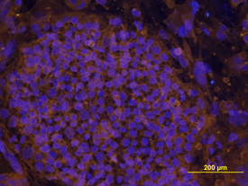
LIN‑41 in BG01V Human Stem Cells. LIN-41 was detected in immersion fixed BG01V human embryonic stem cells using Human LIN-41 Antigen Affinity-purified Polyclonal Antibody (Catalog # AF5104) at 10 µg/mL for 3 hours at room temperature. Cells were stained using the NorthernLights™ 557-conjugated Anti-Sheep IgG Secondary Antibody (yellow; Catalog # NL010) and counterstained with DAPI (blue). View our protocol for Fluorescent ICC Staining of Cells on Coverslips.
 View Larger
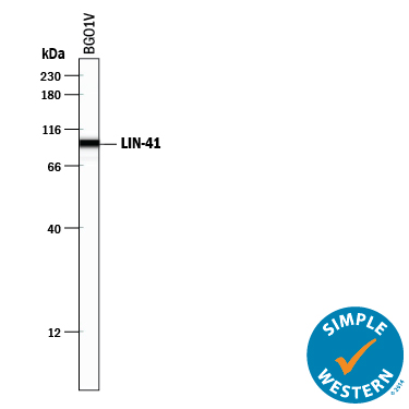
Detection of Human LIN‑41 by Simple WesternTM. Simple Western lane view shows lysates of BG01V human embryonic stem cells, loaded at 0.2 mg/mL. A specific band was detected for LIN-41 at approximately 97 kDa (as indicated) using 10 µg/mL of Sheep Anti-Human LIN-41 Antigen Affinity-purified Polyclonal Antibody (Catalog # AF5104) followed by 1:50 dilution of HRP-conjugated Anti-Sheep IgG Secondary Antibody (Catalog # HAF016). This experiment was conducted under reducing conditions and using the 12-230 kDa separation system.
Reconstitution Calculator
Preparation and Storage
- 12 months from date of receipt, -20 to -70 °C as supplied.
- 1 month, 2 to 8 °C under sterile conditions after reconstitution.
- 6 months, -20 to -70 °C under sterile conditions after reconstitution.
Background: LIN-41
LIN-41 (lineage-41; also TRIM/tripartite motif-containing protein 71) is a 94 kDa (predicted) cytosolic member of the RBCC (RING finger, B-Box Coiled-Coil) family of proteins. It is expressed in developing limbs, DRG and branchial arches and apparently regulates the timed expression of proliferation and differentiation-associated genes. Human LIN-41 is 868 amino acids (aa) in length. It contains a RING-type zinc finger region (aa 12-95), a His-rich segment (aa 146‑157), two B Box zinc fingers that bind DNA (aa 194-314), a coiled-coil motif that binds protein (aa 391-427), one filamin repeat and six NHL repeats that regulate gene translation (aa 593‑868). Over aa 386-868, human LIN-41 is 97%, 95%, and 94% aa identical to canine, mouse, and chicken LIN-41, respectively.
Product Datasheets
Citation for Human LIN-41 Antibody
R&D Systems personnel manually curate a database that contains references using R&D Systems products. The data collected includes not only links to publications in PubMed, but also provides information about sample types, species, and experimental conditions.
1 Citation: Showing 1 - 1
-
T cell activation induces proteasomal degradation of Argonaute and rapid remodeling of the microRNA repertoire.
Authors: Bronevetsky, Yelena, Villarino, Alejandr, Eisley, Christop, Barbeau, Rebecca, Barczak, Andrea J, Heinz, Gitta A, Kremmer, Elisabet, Heissmeyer, Vigo, McManus, Michael, Erle, David J, Rao, Anjana, Ansel, K Mark
J Exp Med, 2013-02-04;210(2):417-32.
Species: Mouse
Sample Types: Whole Cells
Applications: Western Blot
FAQs
No product specific FAQs exist for this product, however you may
View all Antibody FAQsReviews for Human LIN-41 Antibody
There are currently no reviews for this product. Be the first to review Human LIN-41 Antibody and earn rewards!
Have you used Human LIN-41 Antibody?
Submit a review and receive an Amazon gift card.
$25/€18/£15/$25CAN/¥75 Yuan/¥2500 Yen for a review with an image
$10/€7/£6/$10 CAD/¥70 Yuan/¥1110 Yen for a review without an image

