Human MEGF9 Antibody Summary
Ala36-Asn514
Accession # Q9H1U4
Applications
Please Note: Optimal dilutions should be determined by each laboratory for each application. General Protocols are available in the Technical Information section on our website.
Scientific Data
 View Larger
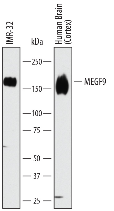
Detection of Human MEGF9 by Western Blot. Western blot shows lysates of IMR-32 human neuroblastoma cell line and human brain (cortex) tissue. PVDF membrane was probed with 1 µg/mL of Sheep Anti-Human MEGF9 Antigen Affinity-purified Polyclonal Antibody (Catalog # AF7768) followed by HRP-conjugated Anti-Sheep IgG Secondary Antibody (Catalog # HAF016). A specific band was detected for MEGF9 at approximately 160 kDa (as indicated). This experiment was conducted under reducing conditions and using Immunoblot Buffer Group 1.
 View Larger
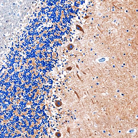
MEGF9 in Human Brain. MEGF9 was detected in immersion fixed paraffin-embedded sections of human brain (cerebellum) using Sheep Anti-Human MEGF9 Antigen Affinity-purified Polyclonal Antibody (Catalog # AF7768) at 1 µg/mL overnight at 4 °C. Before incubation with the primary antibody, tissue was subjected to heat-induced epitope retrieval using Antigen Retrieval Reagent-Basic (Catalog # CTS013). Tissue was stained using the Anti-Sheep HRP-DAB Cell & Tissue Staining Kit (brown; Catalog # CTS019) and counter-stained with hematoxylin (blue). Specific staining was localized to Purkinje neurons. View our protocol for Chromogenic IHC Staining of Paraffin-embedded Tissue Sections.
Reconstitution Calculator
Preparation and Storage
- 12 months from date of receipt, -20 to -70 °C as supplied.
- 1 month, 2 to 8 °C under sterile conditions after reconstitution.
- 6 months, -20 to -70 °C under sterile conditions after reconstitution.
Background: MEGF9
MEGF9 (Multiple EGF‑like domains protein 9; also EGF‑like protein 5) is a 63 kDa (predicted) novel transmembrane glycoprotein that shares some homology to beta ‑chains of laminin. It is expressed by hepatocytes, cerebellar Purkinje cells, Schwann cells, keratinocytes and intestinal epithelium. MEGF9 is suggested to participate in cell motility, and its absence correlates with tumor cell migration. Mature human MEGF9 is 572 amino acids (aa) in length. It is a single span type I transmembrane protein that contains a 484 aa extracellular region (aa 31‑514) plus a 68 aa C‑terminal cytoplasmic domain. The extracellular region possesses a lengthy Pro‑rich region (aa 55‑200), followed by five EGF‑like domains (aa 204‑451). MEGF9 may run at approximately 160 kDa in SDS‑PAGE, suggesting either heavy glycosylation or dimerization. Over aa 36‑514, human MEGF9 shares 76% aa sequence identity with mouse MEGF9.
Product Datasheets
Citation for Human MEGF9 Antibody
R&D Systems personnel manually curate a database that contains references using R&D Systems products. The data collected includes not only links to publications in PubMed, but also provides information about sample types, species, and experimental conditions.
1 Citation: Showing 1 - 1
-
Functionally distinct PI 3-kinase pathways regulate myelination in the peripheral nervous system.
Authors: Heller B, Ghidinelli M, Voelkl J, Einheber S, Smith R, Grund E, Morahan G, Chandler D, Kalaydjieva L, Giancotti F, King R, Fejes-Toth A, Fejes-Toth G, Feltri M, Lang F, Salzer J
J Cell Biol, 2014-03-31;204(7):1219-36.
Species: Mouse, Rat
Sample Types: Whole Tissue
Applications: Bioassay
FAQs
No product specific FAQs exist for this product, however you may
View all Antibody FAQsReviews for Human MEGF9 Antibody
There are currently no reviews for this product. Be the first to review Human MEGF9 Antibody and earn rewards!
Have you used Human MEGF9 Antibody?
Submit a review and receive an Amazon gift card.
$25/€18/£15/$25CAN/¥75 Yuan/¥2500 Yen for a review with an image
$10/€7/£6/$10 CAD/¥70 Yuan/¥1110 Yen for a review without an image

