Human Midkine Antibody Summary
Applications
Please Note: Optimal dilutions should be determined by each laboratory for each application. General Protocols are available in the Technical Information section on our website.
Scientific Data
 View Larger
Detection of Human Midkine by Western Blot. Western blot shows lysates of SH-SY5Y human neuroblastoma cell line. PVDF membrane was probed with 2 µg/mL of Goat Anti-Human Midkine Antigen Affinity-purified Polyclonal Antibody (Catalog # AF-258-PB) followed by HRP-conjugated Anti-Goat IgG Secondary Antibody (HAF017). A specific band was detected for Midkine at approximately 17 kDa (as indicated). This experiment was conducted under reducing conditions and using Immunoblot Buffer Group 1.
 View Larger
Midkine in Human Prostate. Midkine was detected in immersion fixed paraffin-embedded sections of human prostate using Goat Anti-Human Midkine Antigen Affinity-purified Polyclonal Antibody (Catalog # AF-258-PB) at 10 µg/mL overnight at 4 °C. Before incubation with the primary antibody, tissue was subjected to heat-induced epitope retrieval using Antigen Retrieval Reagent-Basic (CTS013). Tissue was stained using the Anti-Goat HRP-DAB Cell & Tissue Staining Kit (brown CTS008) and counterstained with hematoxylin (blue). Specific staining was localized to stromal cell cytoplasm. View our protocol for Chromogenic IHC Staining of Paraffin-embedded Tissue Sections.
 View Larger
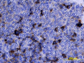
Midkine in Human Ovarian Array. Midkine was detected in immersion fixed paraffin-embedded sections of human ovarian array using Goat Anti-Human Midkine Antigen Affinity-purified Polyclonal Antibody (Catalog # AF-258-PB) at 10 µg/mL overnight at 4 °C. Tissue was stained using the Anti-Goat HRP-DAB Cell & Tissue Staining Kit (brown; CTS008) and counterstained with hematoxylin (blue). View our protocol for Chromogenic IHC Staining of Paraffin-embedded Tissue Sections.
 View Larger
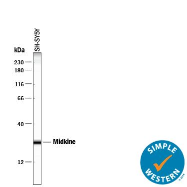
Detection of Human Midkine by Simple WesternTM. Simple Western lane view shows lysates of SH-SY5Y human neuroblastoma cell line, loaded at 0.2 mg/mL. A specific band was detected for Midkine at approximately 26 kDa (as indicated) using 100 µg/mL of Goat Anti-Human Midkine Antigen Affinity-purified Polyclonal Antibody (Catalog # AF-258-PB) followed by 1:50 dilution of HRP-conjugated Anti-Goat IgG Secondary Antibody (HAF109). This experiment was conducted under reducing conditions and using the 12-230 kDa separation system.
 View Larger
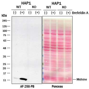
Western Blot Shows Human Midkine Specificity Using Knockout Cell Line. Western blot shows lysates of HAP1 human near-haploid cell line untreated (-) or treated (+) with 3.0 ug/ml Brefeldin A for 18 hr and Midkine knockout HAP1 cell line (KO). Nitrocellulose membrane was probed with 2 µg/mL of Goat Anti-Human Midkine Antigen Affinity-purified Polyclonal Antibody (Catalog # AF-258-PB) followed by HRP-conjugated Anti-Goat IgG Secondary Antibody. A specific band was detected for Midkine at approximately 11 kDa (as indicated) in the parental HAP1 cell line, but is not detectable in knockout HAP1 cell line. The Ponceau stained transfer of the blot is shown. This experiment was conducted under reducing conditions. Image, protocol, and testing courtesy of YCharOS Inc. See ycharos.com for additional details.
 View Larger
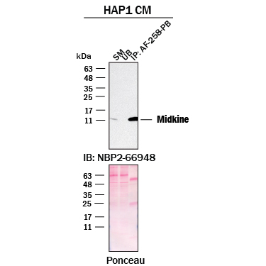
Detection of Midkine by Immunoprecipitation. Immunoprecipitation was performed on concentrated culture media of HAP1 human near-haploid cell line using 2.0 μg of Mouse Anti-Human Midkine Monoclonal Antibody (Catalog # AF-258-PB) pre-coupled to protein G or protein A beads. Immunoprecipitated Midkine was detected with Rabbit Anti-Human Midkine Monoclonal Antibody (NBP2-66948 ) at 1/500. The Ponceau stained transfers of each blot are shown. SM=10% starting material; UB=10% unbound fraction; IP=immunoprecipitated. Image, protocol, and testing courtesy of YCharOS Inc. (ycharos.com).
Reconstitution Calculator
Preparation and Storage
- 12 months from date of receipt, -20 to -70 degreesC as supplied. 1 month, 2 to 8 degreesC under sterile conditions after reconstitution. 6 months, -20 to -70 degreesC under sterile conditions after reconstitution.
Background: Midkine
Midkine (MK) is a 15 kDa heparin-binding molecule originally cloned during a search for genes preferentially transcribed during retinoic acid (RA)-induced differentiation. Midkine belongs to a family of neurotrophic and developmentally-regulated heparin-binding molecules consisting of midkine, pleiotrophin (PTN/HBNF/OSF-1/HNGF-8) and the avian midkine homolog, RI-HB (for retinoic acid-inducible heparin-binding protein). Midkine is a highly basic, nonglycosylated polypeptide that contains five intrachain disulfide bonds. The predicted molecular weight is approximately 13.3 kDa, based on a mature peptide length of 118 amino acid residues in the mouse and 121 amino acid residues in the human. Across species, MK shows 87% identity between the human and murine proteins. Between family members, human MK is approximately 50% identical to human PTN, with conservation of all 10 cysteines. Initial structure-function studies indicate that the C-terminal half of MK contains the principal heparin-binding site plus the molecule’s antigenicity and neurite-promoting sequences; while both the C- and N-termini are necessary for the molecule’s neurotrophic effects. Cells known to produce MK include endothelial cells, fetal astrocytes, renal proximal tubule epithelial cells and Wilms’ (kidney) tumor cells. MK has also been identified in the senile plaques of patients with Alzheimer’s disease. The pattern of expression of midkine during development strongly suggests a role for this factor both in epithelial-mesenchymal interactions and in development of the nervous system.
- Bohlen, P. and I. Kovesdi (1991) Prog. Growth Factor Res. 3:143.
- Muramatsu, T. (1993) Int. J. Dev. Biol. 37:183.
Product Datasheets
Citations for Human Midkine Antibody
R&D Systems personnel manually curate a database that contains references using R&D Systems products. The data collected includes not only links to publications in PubMed, but also provides information about sample types, species, and experimental conditions.
5
Citations: Showing 1 - 5
Filter your results:
Filter by:
-
Midkine Is a Potential Therapeutic Target of Tumorigenesis, Angiogenesis, and Metastasis in Non-Small Cell Lung Cancer
Authors: DH Shin, JY Jo, SH Kim, M Choi, C Han, BK Choi, SS Kim
Cancers (Basel), 2020-08-24;12(9):.
Species: Human, Mouse
Sample Types: Whole Tissue
Applications: IHC -
Role of heparin binding growth factors in nigrostriatal dopamine system development and Parkinson's disease.
Authors: Marchionini DM, Lehrmann E, Chu Y, He B, Sortwell CE, Becker KG, Freed WJ, Kordower JH, Collier TJ
Brain Res., 2007-02-22;1147(0):77-88.
Species: Rat
Sample Types: Tissue Homogenates
Applications: Western Blot -
Neuritogenic activity of chondroitin/dermatan sulfate hybrid chains of embryonic pig brain and their mimicry from shark liver. Involvement of the pleiotrophin and hepatocyte growth factor signaling pathways.
Authors: Li F, Shetty AK, Sugahara K
J. Biol. Chem., 2006-12-04;282(5):2956-66.
Species: Mouse
Sample Types: Whole Cells
Applications: Neutralization -
Regulation of MDK expression in human cancer cells modulates sensitivities to various anticancer drugs: MDK overexpression confers to a multi-drug resistance.
Authors: Kang HC, Kim IJ, Park HW, Jang SG, Ahn SA, Yoon SN, Chang HJ, Yoo BC, Park JG
Cancer Lett., 2006-04-27;247(1):40-7.
Species: Human
Sample Types: Cell Lysates
Applications: Western Blot -
The anti-HIV cytokine midkine binds the cell surface-expressed nucleolin as a low affinity receptor.
Authors: Said EA, Krust B, Nisole S, Svab J, Briand JP, Hovanessian AG
J. Biol. Chem., 2002-07-29;277(40):37492-502.
Species: Human
Sample Types: Whole Cells, Yeast Extract
Applications: ICC, Western Blot
FAQs
No product specific FAQs exist for this product, however you may
View all Antibody FAQsReviews for Human Midkine Antibody
Average Rating: 4 (Based on 1 Review)
Have you used Human Midkine Antibody?
Submit a review and receive an Amazon gift card.
$25/€18/£15/$25CAN/¥75 Yuan/¥2500 Yen for a review with an image
$10/€7/£6/$10 CAD/¥70 Yuan/¥1110 Yen for a review without an image
Filter by:
