Human MMR/CD206 Antibody Summary
Leu19-Lys1383 (Thr399Ala) & (Leu407Phe)
Accession # P22897
Applications
Please Note: Optimal dilutions should be determined by each laboratory for each application. General Protocols are available in the Technical Information section on our website.
Scientific Data
 View Larger
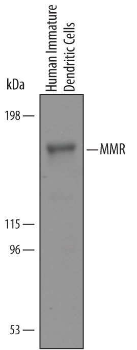
Detection of Human MMR/CD206 by Western Blot. Western blot shows lysates of human immature dendritic cells. PVDF Membrane was probed with 1 µg/mL of Goat Anti-Human MMR/CD206 Antigen Affinity-purified Polyclonal Antibody (Catalog # AF2534) followed by HRP-conjugated Anti-Goat IgG Secondary Antibody (Catalog # HAF019). A specific band was detected for MMR/CD206 at approximately 185 kDa (as indicated). This experiment was conducted under reducing conditions and using Immunoblot Buffer Group 8.
 View Larger
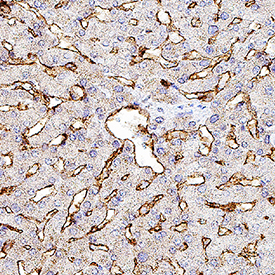
MMR/CD206 in Human Liver. MMR/CD206 was detected in immersion fixed paraffin-embedded sections of human liver using Goat Anti-Human MMR/CD206 Antigen Affinity-purified Polyclonal Antibody (Catalog # AF2534) at 3 µg/mL for 1 hour at room temperature followed by incubation with the Anti-Goat IgG VisUCyte™ HRP Polymer Antibody (VC004). Before incubation with the primary antibody, tissue was subjected to heat-induced epitope retrieval using Antigen Retrieval Reagent-Basic (CTS013). Tissue was stained using DAB (brown) and counterstained with hematoxylin (blue). Specific staining was localized to sinusoids. Staining was performed using our protocol for IHC Staining with VisUCyte HRP Polymer Detection Reagents.
 View Larger
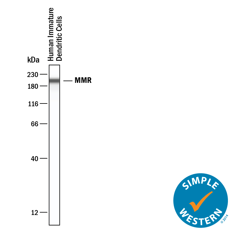
Detection of Human MMR/CD206 by Simple WesternTM. Simple Western lane view shows lysates of human immature dendritic cells, loaded at 0.2 mg/mL. A specific band was detected for MMR/CD206 at approximately 201 kDa (as indicated) using 10 µg/mL of Goat Anti-Human MMR/CD206 Antigen Affinity-purified Polyclonal Antibody (Catalog # AF2534). This experiment was conducted under reducing conditions and using the 12-230 kDa separation system.
Reconstitution Calculator
Preparation and Storage
- 12 months from date of receipt, -20 to -70 °C as supplied.
- 1 month, 2 to 8 °C under sterile conditions after reconstitution.
- 6 months, -20 to -70 °C under sterile conditions after reconstitution.
Background: MMR/CD206
The human Macrophage Mannose Receptor (MMR), also known as CD206 and MRC1 (mannose receptor C, type 1), is a 190 kDa scavenger receptor that is expressed on tissue macrophages, myeloid dendritic cells, and liver and lymphatic endothelial cells (1). It belongs to a family of receptors sharing similar protein structure that also includes DEC205, phospholipase A2 receptor, and Endo180 (2, 3). The human MMR protein is synthesized as a 1456 amino acid (aa) precursor that contains an 18 aa signal sequence, a 1371 aa extracellular region, a 21 aa transmembrane segment and a 46 aa cytoplasmic domain (4). Its extracellular region is composed of an N‑terminal cysteine-rich domain, followed by a single fibronectin type II repeat, and eight C-type lectin carbohydrate recognition domains (CRD) (3, 4). Human to mouse, the extracellular region is 82% aa identical. The cysteine-rich domain mediates recognition of sulfated N‑acetylgalactosamine, which occurs on some extracellular matrix proteins and is the terminal sugar of the unusual oligosaccharides present on pituitary hormones such as lutropin and thyrotropin (5). Several of the CRDs participate in the Ca2+-dependent recognition of carbohydrates showing a preference for branched sugars with terminal mannose, fucose or N‑acetylglucosamine (6). The cytoplasmic domain of MMR includes a tyrosine-based motif for internalization in clathrin-coated vesicles. Once internalized, ligands are released following acidification of phagosomes or endosomes, and the receptor is recycled to the cell surface (3, 7). MMR mediates phagocytosis upon binding to target structures that occur on a variety of pathogenic microorganisms including Gram-negative and Gram-positive bacteria, yeasts, parasites, and mycobacteria. MMR also functions to maintain homeostasis through the endocytosis of potentially harmful glycoproteins associated with inflammation (2, 3).
- East, L. and C. Isake (2002) Biochim. Biophys. Acta 1572:364.
- Chieppa, M. et al. (2003) J. Immunol. 171:4552.
- Figdor, C. et al. (2002) Nat. Rev. Immunol. 2:77.
- Taylor, M. et al. (1990) J. Biol. Chem. 265:12156.
- Leteux, C. et al. (2000) J. Exp. Med. 191:1117.
- Martinez-Pomares, L. et al. (2001) Immunobiology 204:527.
- Feinberg, H. et al. (2000) J. Biol. Chem. 275:21539.
Product Datasheets
Citations for Human MMR/CD206 Antibody
R&D Systems personnel manually curate a database that contains references using R&D Systems products. The data collected includes not only links to publications in PubMed, but also provides information about sample types, species, and experimental conditions.
22
Citations: Showing 1 - 10
Filter your results:
Filter by:
-
Leishmania braziliensis enhances monocyte responses to promote anti-tumor activity
Authors: Dos Santos, JC;Moreno, M;Teufel, LU;Chilibroste, S;Keating, ST;Groh, L;Domínguez-Andrés, J;Williams, DL;Ma, Z;Lowman, DW;Ensley, HE;Novakovic, B;Ribeiro-Dias, F;Netea, MG;Chabalgoity, JA;Joosten, LAB;
Cell reports
Species: Human
Sample Types: Whole Cells
Applications: Functional Assay -
Graph Fourier transform for spatial omics representation and analyses of complex organs
Authors: Ma, Q;Chang, Y;Liu, J;Jiang, Y;Ma, A;Yeo, YY;Guo, Q;McNutt, M;Krull, J;Rodig, S;Barouch, D;Nolan, G;Xu, D;Jiang, S;Li, Z;Liu, B;
Research square
Species: Human
Sample Types: Whole Cells
Applications: Flow Cytometry -
Pro-inflammatory macrophages impair skeletal muscle regeneration in ischemic-damaged limbs by inducing precocious differentiation of satellite cells
Authors: KW Southerlan, Y Xu, DT Peters, X Wei, X Lin, Y Xiang, K Fei, LA Olivere, JM Morowitz, J Otto, Q Dai, CD Kontos, Y Diao
bioRxiv : the preprint server for biology, 2023-04-03;0(0):.
Species: Human
Sample Types: Whole Tissue
Applications: IHC -
CD206+ tumor-associated macrophages interact with CD4+ tumor-infiltrating lymphocytes and predict adverse patient outcome in human laryngeal squamous cell carcinoma
Authors: Y Heng, X Zhu, H Lin, M Jingyu, X Ding, L Tao, L Lu
Journal of Translational Medicine, 2023-03-03;21(1):167.
Species: Human
Sample Types: Whole Tissue
Applications: IHC -
An immunoPET probe to SARS-CoV-2 reveals early infection of the male genital tract in rhesus macaques
Authors: P Madden, Y Thomas, R Blair, S Samer, M Doyle, C Midkiff, L Doyle-Meye, M Becker, S Arif, M McRaven, L Simons, A Carias, E Martinelli, R Lorenzo-Re, J Hultquist, F Villinger, R Veazey, T Hope
Research square, 2022-04-08;0(0):.
Species: Primate - Macaca mulatta (Rhesus Macaque)
Sample Types: Whole Tissue
Applications: Immunohistochemistry -
Macrophages expressing uncoupling protein 1 increase in adipose tissue in response to cold in humans
Authors: BS Finlin, H Memetimin, AL Confides, B Zhu, PM Westgate, EE Dupont-Ver, PA Kern
Scientific Reports, 2021-12-08;11(1):23598.
Species: Human
Sample Types: Whole Tissue
Applications: IHC -
Interaction between Macrophages and Human Mesenchymal Stromal Cells Derived from Bone Marrow and Wharton's Jelly-A Comparative Study
Authors: M Dymowska, A Aksamit, K Zielniok, M Kniotek, B Kaleta, A Roszczyk, M Zych, F Dabrowski, L Paczek, A Burdzinska
Pharmaceutics, 2021-11-01;13(11):.
Species: Human
Sample Types: Whole Cells
Applications: ICC -
Partial Ablation of Non-Myogenic Progenitor Cells as a Therapeutic Approach to Duchenne Muscular Dystrophy
Authors: Z Gao, A Lu, AC Daquinag, Y Yu, M Huard, C Tseng, X Gao, J Huard, MG Kolonin
Biomolecules, 2021-10-15;11(10):.
Species: Mouse
Sample Types: Whole Tissue
Applications: IHC -
Opposing functions of ?-arrestin 1 and 2 in Parkinson's disease via microglia inflammation and Nprl3
Authors: Fang Y, Jiang Q, Li S et al.
Cell Death & Differentiation
-
The Metalloproteinase ADAMTS5 Is Expressed by Interstitial Inflammatory Cells in IgA Nephropathy and Is Proteolytically Active on the Kidney Matrix
Authors: S Taylor, M Whitfield, J Barratt, A Didangelos
J. Immunol., 2020-09-11;0(0):.
Species: Human
Sample Types: Whole Tissue
Applications: IHC -
Correlations of Calf Muscle Macrophage Content With Muscle Properties and Walking Performance in Peripheral Artery Disease
Authors: K Kosmac, M Gonzalez-F, MM McDermott, SH White, RG Walton, RL Sufit, L Tian, L Li, MR Kibbe, MH Criqui, JM Guralnik, T S Polonsky, C Leeuwenbur, L Ferrucci, CA Peterson
J Am Heart Assoc, 2020-05-09;9(10):e015929.
Species: Human
Sample Types: Whole Tissue
Applications: IHC -
Skewed macrophage polarization in aging skeletal muscle
Authors: CY Cui, RK Driscoll, Y Piao, CW Chia, M Gorospe, L Ferrucci
Aging Cell, 2019-09-02;0(0):e13032.
Species: Human
Sample Types: Whole Tissue
Applications: IHC-Fr -
Markers of adipose tissue inflammation are transiently elevated during intermittent fasting in women who are overweight or obese
Authors: B Liu, AT Hutchison, CH Thompson, K Lange, LK Heilbronn
Obes Res Clin Pract, 2019-07-11;0(0):.
Species: Human
Sample Types: Whole Tissue
Applications: IHC-P -
The macrophage-related biomarkers sCD163 and sCD206 are released by different shedding mechanisms
Authors: MC Nielsen, MN Andersen, N Rittig, S Rødgaard-H, H Grønbaek, SK Moestrup, HJ Møller, A Etzerodt
J. Leukoc. Biol., 2019-06-26;0(0):.
Species: Human
Sample Types: Cell Culture Supernates
Applications: ELISA Capture -
Interaction with tumor?associated macrophages promotes PRL?3?induced invasion of colorectal cancer cells via MAPK pathway?induced EMT and NF??B signaling?induced angiogenesis
Authors: T Zhang, L Liu, W Lai, Y Zeng, H Xu, Q Lan, P Su, Z Chu
Oncol. Rep., 2019-03-07;0(0):.
Species: Human
Sample Types: Whole Cells
Applications: Flow Cytometry -
A glucose-responsive insulin therapy protects animals against hypoglycemia
Authors: R Yang, M Wu, S Lin, RP Nargund, X Li, T Kelly, L Yan, G Dai, Y Qian, Q Dallas-Yan, PA Fischer, Y Cui, X Shen, P Huo, DD Feng, MD Erion, DE Kelley, J Mu
JCI Insight, 2018-01-11;3(1):.
Applications: Bioassay -
Regulatory role of cytosolic phospholipase A2 alpha in the induction of CD40 in microglia
Authors: YF Malada-Ede, N Hadad, R Levy
J Neuroinflammation, 2017-02-10;14(1):33.
Species: Mouse
Sample Types: Whole Tissue
Applications: IHC -
FITC Conjugation Markedly Enhances Hepatic Clearance of N-Formyl Peptides
PLoS ONE, 2016-08-05;11(8):e0160602.
Species: Human, Mouse
Sample Types: Whole Tissue
Applications: IHC -
MicroRNA29a regulates IL-33-mediated tissue remodelling in tendon disease.
Authors: Millar N, Gilchrist D, Akbar M, Reilly J, Kerr S, Campbell A, Murrell G, Liew F, Kurowska-Stolarska M, McInnes I
Nat Commun, 2015-04-10;6(0):6774.
Species: Human, Mouse
Sample Types: Whole Tissue
Applications: IHC -
Pioglitazone treatment reduces adipose tissue inflammation through reduction of mast cell and macrophage number and by improving vascularity.
Authors: Spencer M, Yang L, Adu A, Finlin B, Zhu B, Shipp L, Rasouli N, Peterson C, Kern P
PLoS ONE, 2014-07-10;9(7):e102190.
Species: Human
Sample Types: Whole Tissue
Applications: IHC-P -
Targeting the ANG2/TIE2 axis inhibits tumor growth and metastasis by impairing angiogenesis and disabling rebounds of proangiogenic myeloid cells.
Authors: Mazzieri R, Pucci F, Moi D, Zonari E, Ranghetti A, Berti A, Politi LS, Gentner B, Brown JL, Naldini L, De Palma M
Cancer Cell, 2011-04-12;19(4):512-26.
Species: Mouse
Sample Types: Whole Tissue
Applications: IHC -
Acid fibroblast growth factor and peripheral nerve grafts regulate Th2 cytokine expression, macrophage activation, polyamine synthesis, and neurotrophin expression in transected rat spinal cords.
Authors: Kuo HS, Tsai MJ, Huang MC
J. Neurosci., 2011-03-16;31(11):4137-47.
Species: Rat
Sample Types: Whole Tissue
Applications: IHC
FAQs
No product specific FAQs exist for this product, however you may
View all Antibody FAQsReviews for Human MMR/CD206 Antibody
Average Rating: 3 (Based on 1 Review)
Have you used Human MMR/CD206 Antibody?
Submit a review and receive an Amazon gift card.
$25/€18/£15/$25CAN/¥75 Yuan/¥2500 Yen for a review with an image
$10/€7/£6/$10 CAD/¥70 Yuan/¥1110 Yen for a review without an image
Filter by:
I used this antibody for developing a sandwich ELISA in combination with mAb (cat.MAB25341) and protein (cat.2534-MR). This combination gives good standard curve but unfortunately did not detect any MMR in our samples.


