Human/Mouse BID Antibody Summary
Met1-Asp195
Accession # P70444
Applications
Please Note: Optimal dilutions should be determined by each laboratory for each application. General Protocols are available in the Technical Information section on our website.
Scientific Data
 View Larger
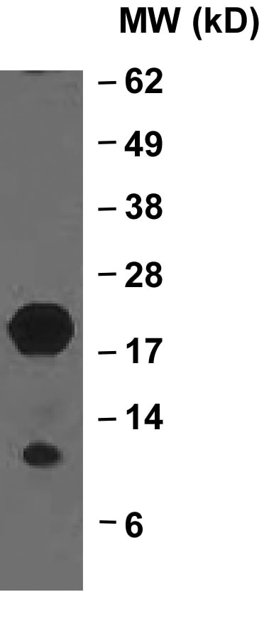
Detection of Human and Mouse BID by Western Blot. Western blot shows lysates of Recombinant Mouse BID and Recombinant Human BID untreated (-) or treated (+) with Caspase-8. PVDF membrane was probed with 1 µg/mL Rat Anti-Human/Mouse BID Monoclonal Antibody (Catalog # MAB860) followed by HRP-conjugated Anti-Rat IgG Secondary Antibody (Catalog # HAF005). Specific bands for BID were detected at approximately 12 kDa and 20 kDa (as indicated). This experiment was conducted under reducing conditions and using Immunoblot Buffer Group 4.
 View Larger
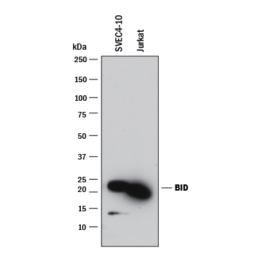
Detection of Human and Mouse BID by Western Blot. Western blot shows lysates of SVEC4-10 mouse vascular endothelial cell line and Jurkat human acute T cell leukemia cell line. PVDF membrane was probed with 1 µg/mL of Rat Anti-Human/Mouse BID Monoclonal Antibody (Catalog # MAB860) followed by HRP-conjugated Anti-Rat IgG Secondary Antibody (Catalog # HAF005). A specific band was detected for BID at approximately 20 kDa (as indicated). This experiment was conducted under reducing conditions and using Immunoblot Buffer Group 1.
Reconstitution Calculator
Preparation and Storage
- 12 months from date of receipt, -20 to -70 °C as supplied.
- 1 month, 2 to 8 °C under sterile conditions after reconstitution.
- 6 months, -20 to -70 °C under sterile conditions after reconstitution.
Background: BID
BID is a member of the Bcl-2 family of proteins that regulates outer mitochondrial membrane permeability. BID is cytosolic in healthy cells but upon delivery of an apoptotic signal, BID is cleaved by Caspase-8. The cleaved form is translocated to the mitochondria outer membrane where it binds to BAK and the resulting complex causes altered mitochondrial membrane permeability.
Product Datasheets
Citations for Human/Mouse BID Antibody
R&D Systems personnel manually curate a database that contains references using R&D Systems products. The data collected includes not only links to publications in PubMed, but also provides information about sample types, species, and experimental conditions.
8
Citations: Showing 1 - 8
Filter your results:
Filter by:
-
Apoptotic signaling clears engineered Salmonella in an organ-specific manner
Authors: Abele, TJ;Billman, ZP;Li, L;Harvest, CK;Bryan, AK;Magalski, GR;Lopez, JP;Larson, HN;Yin, XM;Miao, EA;
bioRxiv : the preprint server for biology
Species: Mouse, Transgenic Mouse
Sample Types: Cell Fraction, Cell Lysates
Applications: Western Blot -
LPS-induced inflammation desensitizes hepatocytes to Fas-induced apoptosis through Stat3 activation-The effect can be reversed by ruxolitinib
Authors: A Markotic, D Flegar, D Grcevic, A Sucur, H Lalic, P Turcic, N Kovacic, N Lukac, D Pravdic, K Vukojevic, I Cavar, T Kelava
J. Cell. Mol. Med., 2020-02-05;0(0):.
Species: Mouse
Sample Types: Tissue Lysates
Applications: Western Blot -
Impact of caspase-1/11, -3, -7, or IL-1?/IL-18 deficiency on rabies virus-induced macrophage cell death and onset of disease
Authors: E Kip, F Nazé, V Suin, T Vanden Ber, A Francart, S Lamoral, P Vandenabee, R Beyaert, S Van Gucht, M Kalai
Cell Death Discov, 2017-03-06;3(0):17012.
Species: Mouse
Sample Types: Cell Lysates
Applications: Western Blot -
Sensing cytosolic RpsL by macrophages induces lysosomal cell death and termination of bacterial infection.
Authors: Zhu, Wenhan, Tao, Lili, Quick, Marsha L, Joyce, Johanna, Qu, Jie-Ming, Luo, Zhao-Qin
PLoS Pathog, 2015-03-04;11(3):e1004704.
Species: Mouse
Sample Types: Cell Lysates
Applications: Western Blot -
The PI3K regulatory subunits p55alpha and p50alpha regulate cell death in vivo.
Authors: Pensa S, Neoh K, Resemann H, Kreuzaler P, Abell K, Clarke N, Reinheckel T, Kahn C, Watson C
Cell Death Differ, 2014-06-06;21(9):1442-50.
Species: Mouse
Sample Types: Cell Lysates
Applications: Western Blot -
BH3-only protein Bid is dispensable for seizure-induced neuronal death and the associated nuclear accumulation of apoptosis-inducing factor.
Authors: Engel T, Caballero-Caballero A, Schindler CK, Plesnila N, Strasser A, Prehn JH, Henshall DC
J. Neurochem., 2010-07-30;115(1):92-101.
Species: Mouse
Sample Types: Tissue Homogenates, Whole Tissue
Applications: IHC-Fr, Western Blot -
Activation of the extrinsic caspase pathway in cultured cortical neurons requires p53-mediated down-regulation of the X-linked inhibitor of apoptosis protein to induce apoptosis.
Authors: Tun C, Guo W, Nguyen H, Yun B, Libby RT, Morrison RS, Garden GA
J. Neurochem., 2007-05-04;102(4):1206-19.
Species: Mouse
Sample Types: Cell Lysates
Applications: Western Blot -
Proteolysis of HIP during apoptosis occurs within a region similar to the BID loop.
Authors: Caruso JA, Reiners JJ
Apoptosis, 2006-11-01;11(11):1877-85.
Species: Mouse
Sample Types: Recombinant Protein
Applications: Western Blot
FAQs
No product specific FAQs exist for this product, however you may
View all Antibody FAQsReviews for Human/Mouse BID Antibody
Average Rating: 4.3 (Based on 3 Reviews)
Have you used Human/Mouse BID Antibody?
Submit a review and receive an Amazon gift card.
$25/€18/£15/$25CAN/¥75 Yuan/¥2500 Yen for a review with an image
$10/€7/£6/$10 CAD/¥70 Yuan/¥1110 Yen for a review without an image
Filter by:
Could only detect the pro form in my samples.


