Human/Mouse c-Myc Antibody Summary
Arg66-Asp201
Accession # P01106
Applications
Please Note: Optimal dilutions should be determined by each laboratory for each application. General Protocols are available in the Technical Information section on our website.
Scientific Data
 View Larger
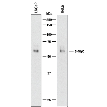
Detection of Human c-Myc by Western Blot. Western blot shows lysates of LNCaP human prostate cancer cell line and HeLa human cervical epithelial carcinoma cell line. PVDF membrane was probed with 0.5 µg/mL of Goat Anti-Human/Mouse c-Myc Antigen Affinity-purified Polyclonal Antibody (Catalog # AF3696) followed by HRP-conjugated Anti-Goat IgG Secondary Antibody (Catalog # HAF017). A specific band was detected for c-Myc at approximately 56 kDa (as indicated). This experiment was conducted under reducing conditions and using Immunoblot Buffer Group 1.
 View Larger
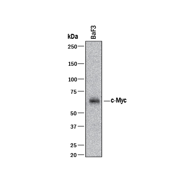
Detection of Mouse c‑Myc by Western Blot. Western blot shows lysates of BaF3 mouse pro-B cell line. PVDF membrane was probed with 0.5 µg/mL of Goat Anti-Human/Mouse c-Myc Antigen Affinity-purified Polyclonal Antibody (Catalog # AF3696) followed by HRP-conjugated Anti-Goat IgG Secondary Antibody (Catalog # HAF017). A specific band was detected for c-Myc at approximately 56 kDa (as indicated). This experiment was conducted under reducing conditions and using Immunoblot Buffer Group 1.
 View Larger
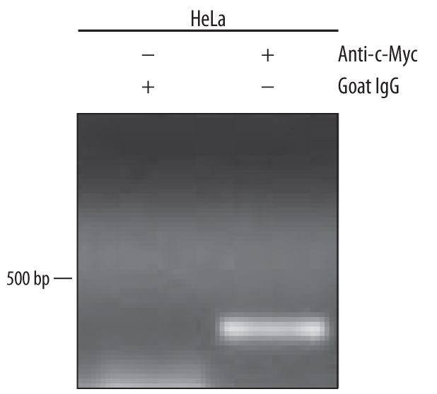
Detection of c‑Myc-regulated Genes by Chromatin Immunoprecipitation. HeLa human cervical epithelial carcinoma cell line was fixed using formaldehyde, resuspended in lysis buffer, and sonicated to shear chromatin. c-Myc/DNA complexes were immunoprecipitated using 5 µg Goat Anti-Human/Mouse c-Myc Antigen Affinity-purified Polyclonal Antibody (Catalog # AF3696) or control antibody (Catalog # AB-108-C) for 15 minutes in an ultrasonic bath, followed by Biotinylated Anti-Goat IgG Secondary Antibody (Catalog # BAF109). Immunocomplexes were captured using 50 µL of MagCellect Streptavidin Ferrofluid (Catalog # MAG999) and DNA was purified using chelating resin solution. Thep21promoter was detected by standard PCR.
 View Larger
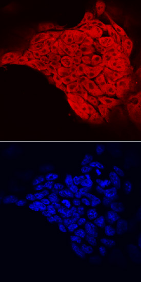
c‑Myc in D3 Mouse Stem Cells. c-Myc was detected in immersion fixed D3 mouse embryonic stem cell line using Goat Anti-Human/Mouse c-Myc Antigen Affinity-purified Polyclonal Antibody (Catalog # AF3696) at 10 µg/mL for 3 hours at room temperature. Cells were stained using the NorthernLights™ 557-conjugated Anti-Goat IgG Secondary Antibody (red, upper panel; Catalog # NL001) and counterstained with DAPI (blue, lower panel). Specific staining was localized to nuclei and cytoplasm. View our protocol for Fluorescent ICC Staining of Cells on Coverslips.
 View Larger
c‑Myc in BG01V Human Stem Cells. c-Myc was detected in immersion fixed BG01V human embryonic stem cells using 10 µg/mL Goat Anti-Human/Mouse c-Myc Antigen Affinity-purified Polyclonal Antibody (Catalog # AF3696) for 3 hours at room temperature. Cells were stained with the NorthernLights™ 557-conjugated Anti-Goat IgG Secondary Antibody (red; Catalog # NL001) and counterstained with DAPI (blue). View our protocol for Fluorescent ICC Staining of Cells on Coverslips.
 View Larger
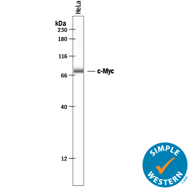
Detection of Human c‑Myc by Simple WesternTM. Simple Western lane view shows lysates of HeLa human cervical epithelial carcinoma cell line, loaded at 0.2 mg/mL. A specific band was detected for c-Myc at approximately 78 kDa (as indicated) using 20 µg/mL of Goat Anti-Human/Mouse c-Myc Antigen Affinity-purified Polyclonal Antibody (Catalog # AF3696) followed by 1:50 dilution of HRP-conjugated Anti-Goat IgG Secondary Antibody (Catalog # HAF109). This experiment was conducted under reducing conditions and using the 12-230 kDa separation system.
 View Larger
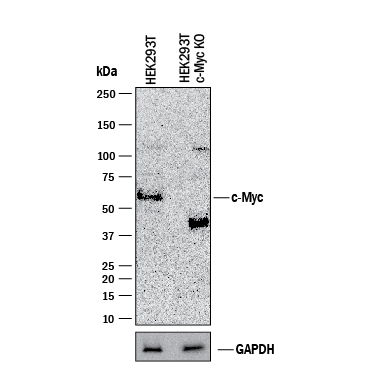
Western Blot Shows Human c‑Myc Specificity by Using Knockout Cell Line. Western blot shows lysates of HEK293T human embryonic kidney parental cell line and c-Myc knockout HEK293T cell line (KO). PVDF membrane was probed with 0.5 µg/mL of Goat Anti-Human/Mouse c-Myc Antigen Affinity-purified Polyclonal Antibody (Catalog # AF3696) followed by HRP-conjugated Anti-Goat IgG Secondary Antibody (Catalog # HAF017). A specific band was detected for c-Myc at approximately 52 kDa (as indicated) in the parental HEK293T cell line, but is not detectable in knockout HEK293T cell line. GAPDH (Catalog # AF5718) is shown as a loading control. This experiment was conducted under reducing conditions and using Immunoblot Buffer Group 1.
Reconstitution Calculator
Preparation and Storage
- 12 months from date of receipt, -20 to -70 °C as supplied.
- 1 month, 2 to 8 °C under sterile conditions after reconstitution.
- 6 months, -20 to -70 °C under sterile conditions after reconstitution.
Background: c-Myc
Human c-Myc is a 439 amino acid transcription factor with a bHLH/LZ (basic Helix-Loop-Helix, Leucine Zipper) domain. c-Myc DNA-binding and transcription function is achieved upon heterodimerization with its partner Max. c-Myc is often over-expressed and mutated in hematopoietic tumors. Mutations frequently result in truncations that remove the transactivation region or in the bHLH/LZ domain required for association with Max and DNA. Over the region used as immunogen, human c-Myc is 92% identical to the rat and mouse c-Myc proteins.
Product Datasheets
Citations for Human/Mouse c-Myc Antibody
R&D Systems personnel manually curate a database that contains references using R&D Systems products. The data collected includes not only links to publications in PubMed, but also provides information about sample types, species, and experimental conditions.
5
Citations: Showing 1 - 5
Filter your results:
Filter by:
-
SCARB2 drives hepatocellular carcinoma tumor initiating cells via enhanced MYC transcriptional activity
Authors: Wang F, Gao Y, Xue S et al.
Nat Commun
-
Chaetocin Promotes Osteogenic Differentiation via Modulating Wnt/Beta-Catenin Signaling in Mesenchymal Stem Cells
Authors: Y Liang, X Liu, R Zhou, D Song, YZ Jiang, W Xue
Stem Cells International, 2021-02-06;2021(0):8888416.
Species: Mouse
Sample Types: Cell Lysates
Applications: Western Blot -
LncRNA SNHG6 regulating Hedgehog signaling pathway and affecting the biological function of gallbladder carcinoma cells through targeting miR-26b-5p
Authors: XF Liu, K Wang, HC Du
Eur Rev Med Pharmacol Sci, 2020-07-01;24(14):7598-7611.
Species: Human
Sample Types: Cell Lysates
Applications: Western Blot -
Lysine-Specific Demethylase 1 (LSD1/KDM1A) Is a Novel Target Gene of c-Myc
Authors: M Nagasaka, K Tsuzuki, Y Ozeki, M Tokugawa, N Ohoka, Y Inoue, H Hayashi
Biol. Pharm. Bull., 2019-01-01;42(3):481-488.
Species: Human
Sample Types: Cell Lysates
Applications: Western Blot -
Convergence of cMyc and beta-catenin on Tcf7l1 enables endoderm specification.
Authors: Morrison G, Scognamiglio R, Trumpp A, Smith A
EMBO J, 2015-12-16;35(3):356-68.
Species: Mouse
Sample Types: Cell Lysates
Applications: ChIP
FAQs
No product specific FAQs exist for this product, however you may
View all Antibody FAQsReviews for Human/Mouse c-Myc Antibody
Average Rating: 5 (Based on 1 Review)
Have you used Human/Mouse c-Myc Antibody?
Submit a review and receive an Amazon gift card.
$25/€18/£15/$25CAN/¥75 Yuan/¥2500 Yen for a review with an image
$10/€7/£6/$10 CAD/¥70 Yuan/¥1110 Yen for a review without an image
Filter by:


